
Dear Visitor! We are carrying out maintenance this weekend, so our website may be slow or briefly unavailable at times. Thank you for your patience!
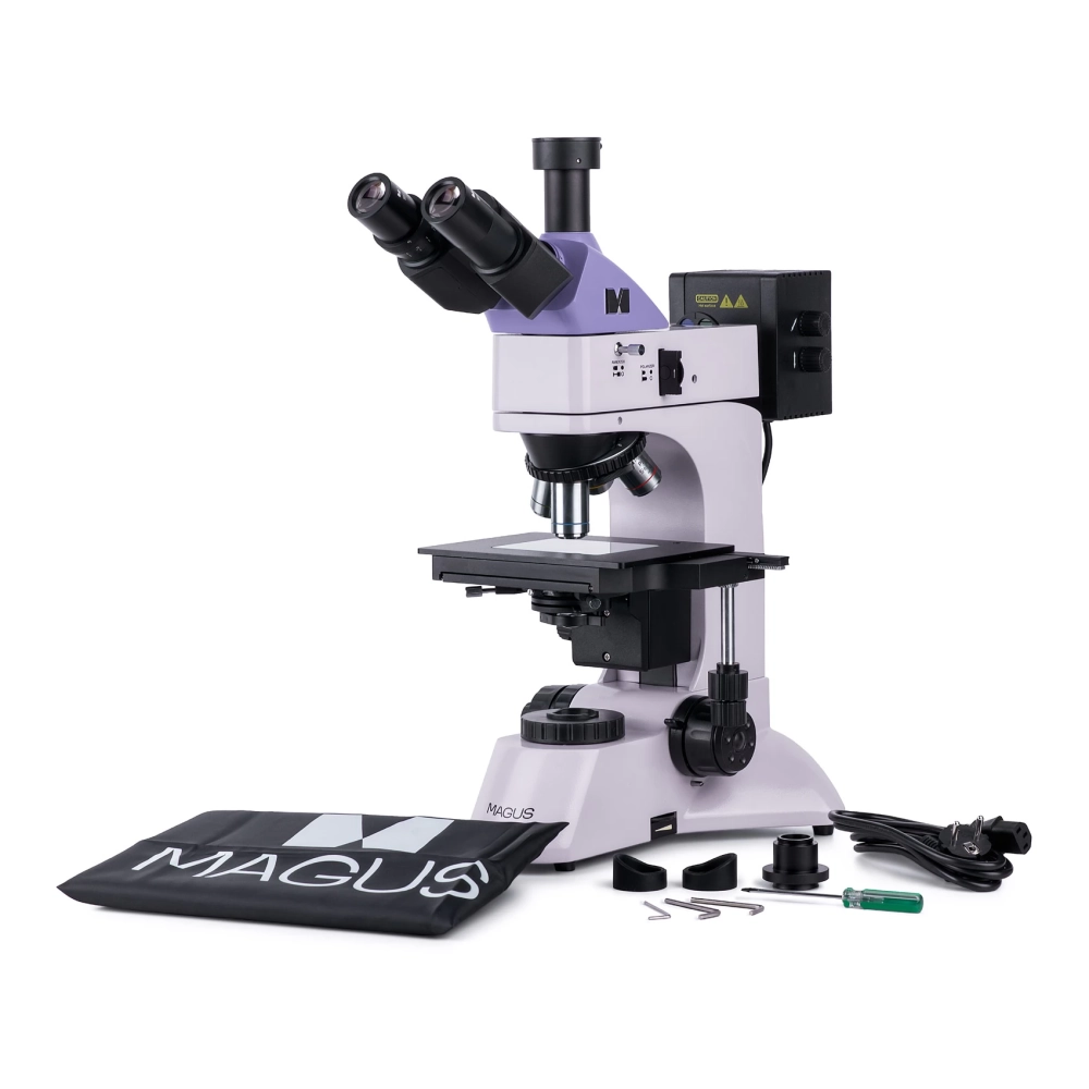
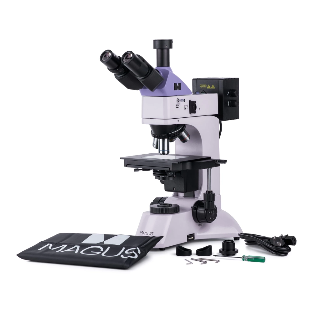
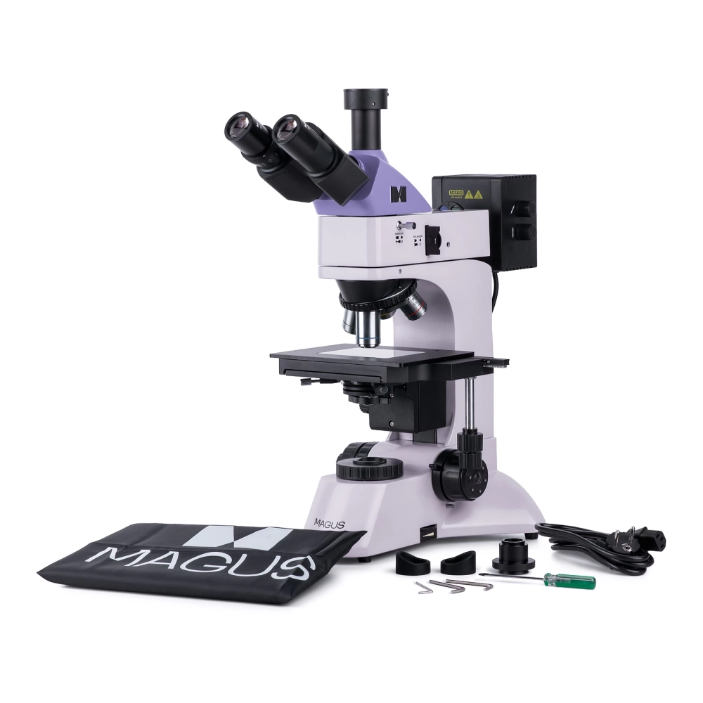
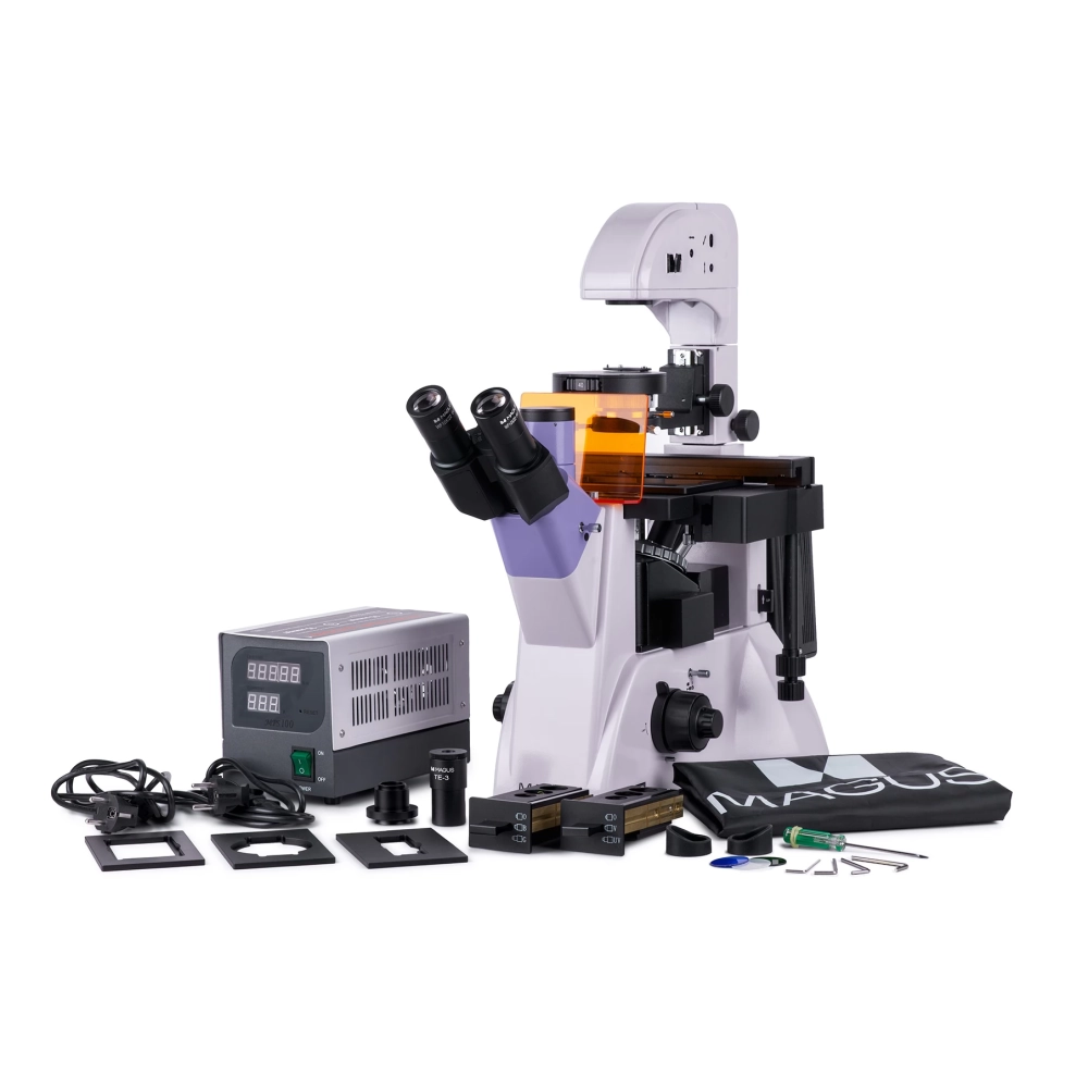
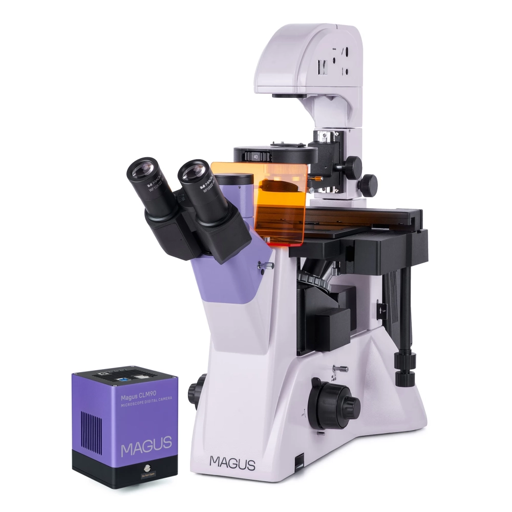
MAGUS Lum VD500 fluoreszcens reverse digital microscope
Product description
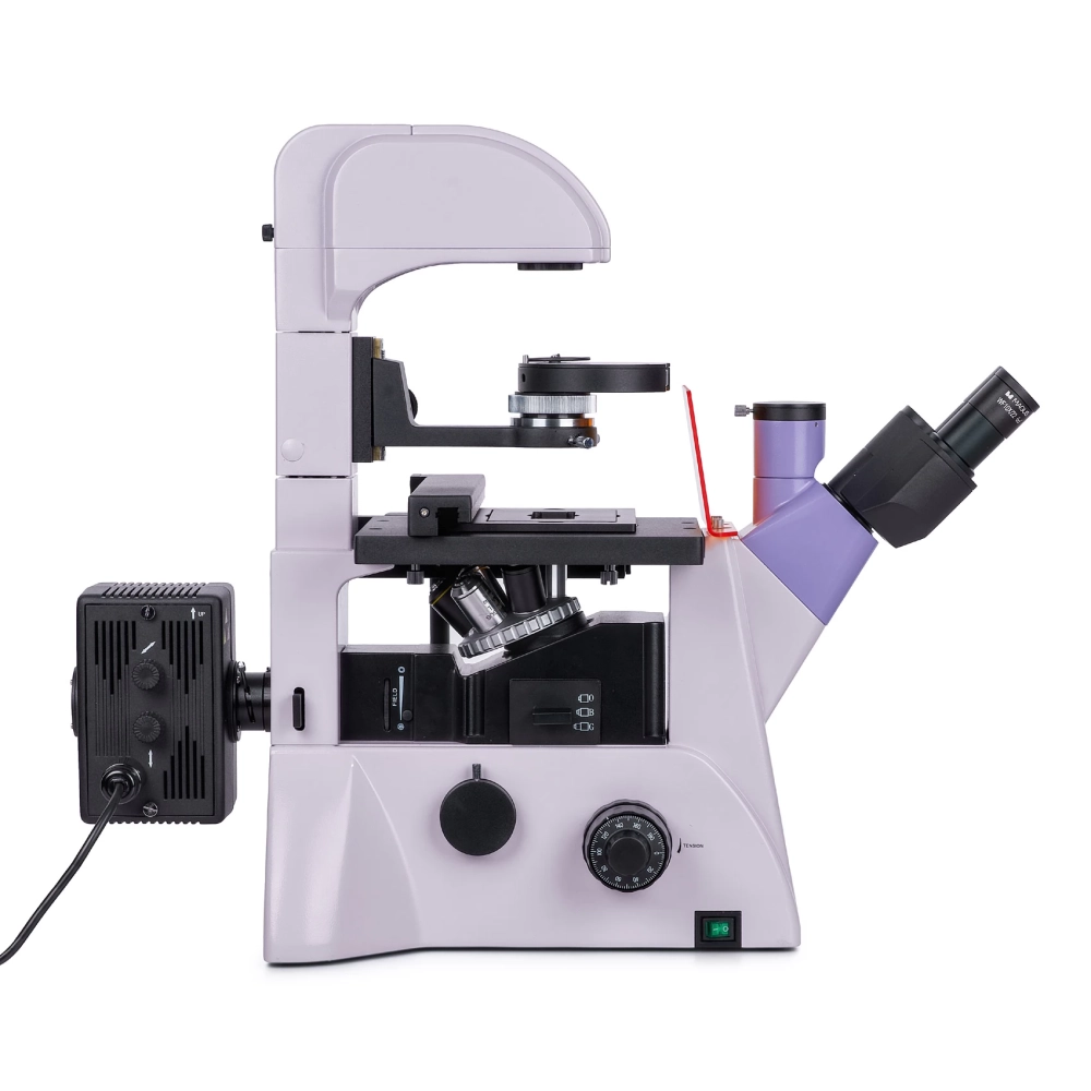
The MAGUS Lum V500 fluorescence inverted microscope is designed to observe samples in laboratory vessels up to 55 mm high and up to 1.2 mm bottom thick. If the lighting stand is tilted, it is possible to use larger laboratory vessels of up to 165 mm. The samples can be observed in reflected light (fluorescence microscopy technique) and in transmitted light (bright field and phase contrast microscopy technique). The main fields of application of the microscope are: medicine, pharmacy, biochemistry, epidemiology, etc.
Digital camera
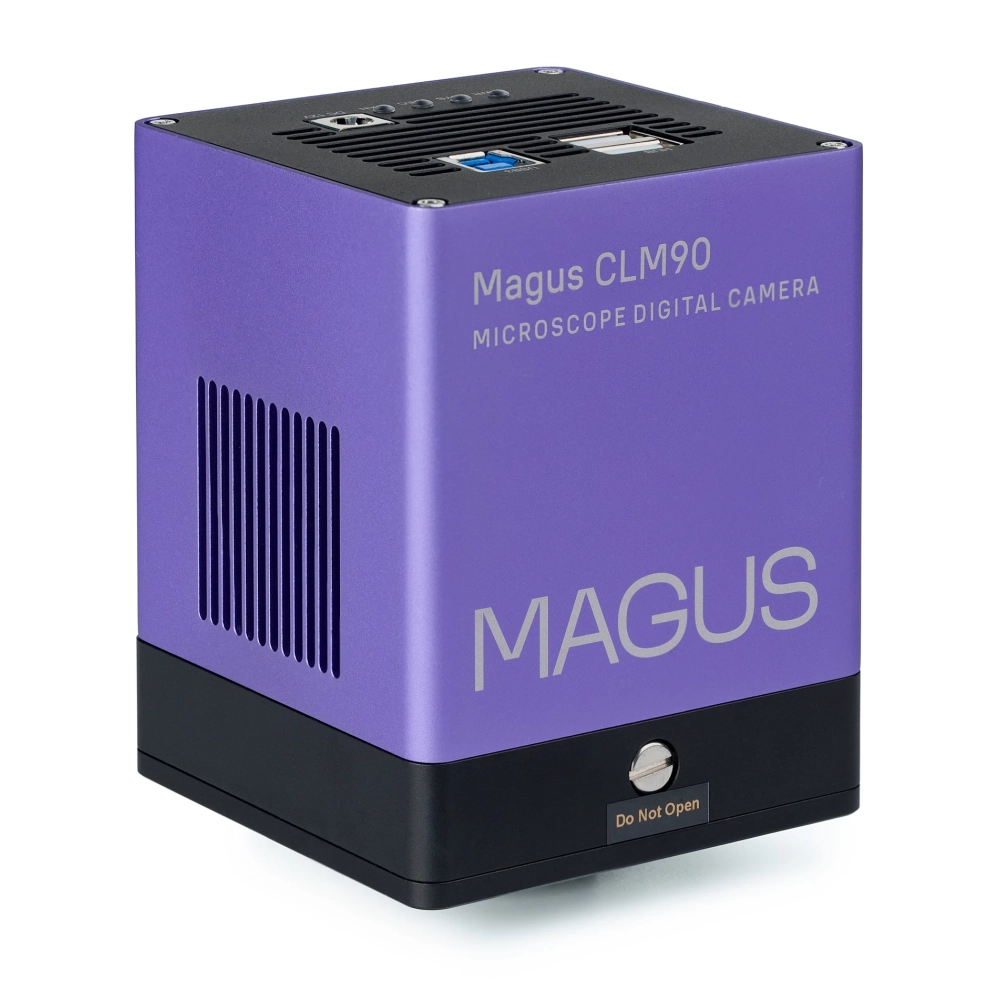
The Magus CLM90 is a digital camera designed for fluorescence and dark field microscopy techniques.
The camera has a 7.1 MP sensor and produces realistic images with a resolution of 3200x2200 pixels. The camera should be used with 4x, 10x, 20x and 40x lenses. When working with low magnification lenses, the camera helps you see the finer details.
Video is recorded at 51.3 frames per second (fps) or 133.8 frames per second, depending on the resolution. The videos are stutter-free, the transition between frames is smooth and smooth. The movement of the sample can be tracked in real time without delay. Moving objects can be easily observed using the camera, which is also suitable for classroom presentations.
The camera has a USB 3.0 interface. The data transfer rate is 10 times faster than USB 2.0 cameras. The high-speed camera is recommended for professional laboratories, research and university education.
Optical elements
The microscope has a trinocular head and a three-eye tube. The microscope has a side camera connector. This design allows you to mount both a digital camera and a monitor on the microscope at the same time. The beam distribution on the three-lens head part is 80:20, on the frame – 100:0/0:100. The eyepiece tubes can be rotated 180°, so the user can adjust the eye distance for himself.
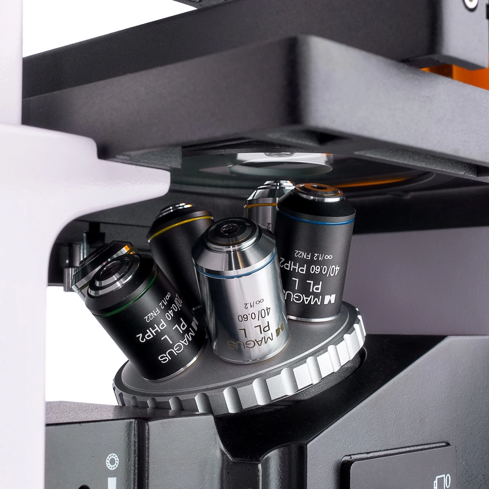
The rotating objective lens holder is attached to the stand under the stage. It has six free openings for attaching lenses. Three phase-contrast lenses and three fluo- and bright-field lenses are included in the package. Flat achromatic objectives with a long working distance for laboratory dishes with a bottom thickness of 1.2 mm.
Illumination
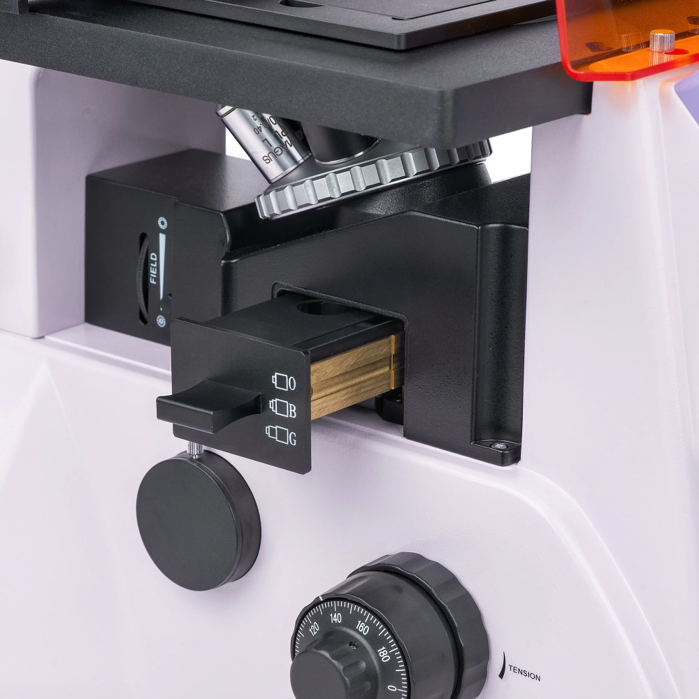
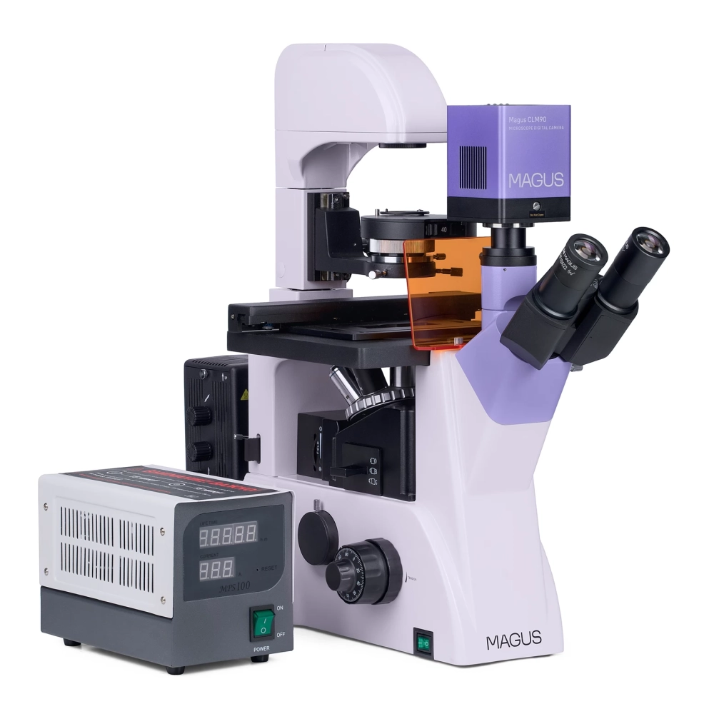
A 100 W mercury vapor lamp serves as a reflective source. In order to reduce the risk of overheating and better heat dissipation during long operation, it is housed in a lamp housing. The lamp housing has a wide range of wavelengths and peak values, which makes it convenient to work with different fluorescent dyes. Fluorescent filters: ultraviolet (UV), violet (V), blue (B) and green (G). The mercury lamp shines brightly, provides good visibility and can be easily replaced if necessary.
The transmitted light is provided by a 30 W halogen lamp. It shines brightly, its light spectrum is warm. Therefore, observation with each lens is effective and comfortable for the eyes. The microscope has a four-hole phase-contrast condenser: One is used for the bright field, and the other three are for the 10x, 20x and 40x phase-contrast objectives. The phase rings can be centered. By rotating the disc, you can switch between bright field of view and phase contrast. It is convenient for research, as it saves time during work.
The Köhler illumination method for transmitted and reflected light. Using this method, we can get a clear, sharp, high-resolution image, without artifacts and darkening of the edges.
Stage and focusing mechanism
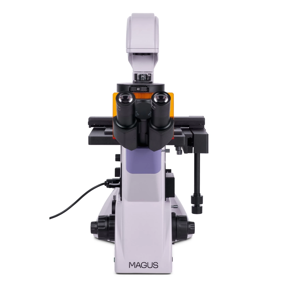
The specimen table is fixed with a mechanism for moving laboratory vessels. The laboratory vessel can be moved horizontally in two mutually perpendicular directions. Its movement is precise and delicate, and it has four pot holders for storing pots of different sizes. The focus of the microscope can be adjusted with coarse and fine focusing buttons. The coarse focus has a lock button and is adjustable in tension. The focus buttons are located coaxially on both sides of the frame.
- For brightfield microscopes with 4x, 10x, 20x or 40x objectives
- 7.1 MP resolution: when observing with low magnification lenses, the camera helps you to see the finer details
- 51.3 frames/sec (fps) with a resolution of 3200x2200 pixels, and 113.8 frames/sec with a resolution of 1584x1100 pixels for observing moving objects, recording videos, and moving the product without stuttering or delay
- The Peltier element reduces the temperature of the sensor during longer operation and eliminates thermal noise, so longer shutter speeds can be achieved when taking photos
- Global lock for quick reading of the signal from the sensor, for greater brightness of the image, and for avoiding incorrect optical effects when observing moving objects
- The SONY Exmor monochrome CMOS backlit sensor provides low noise and high light sensitivity even in low light conditions. It is cleaner, brighter and more saturated
NOTEBOOK SEARCH

OPINIONS
0.0
