
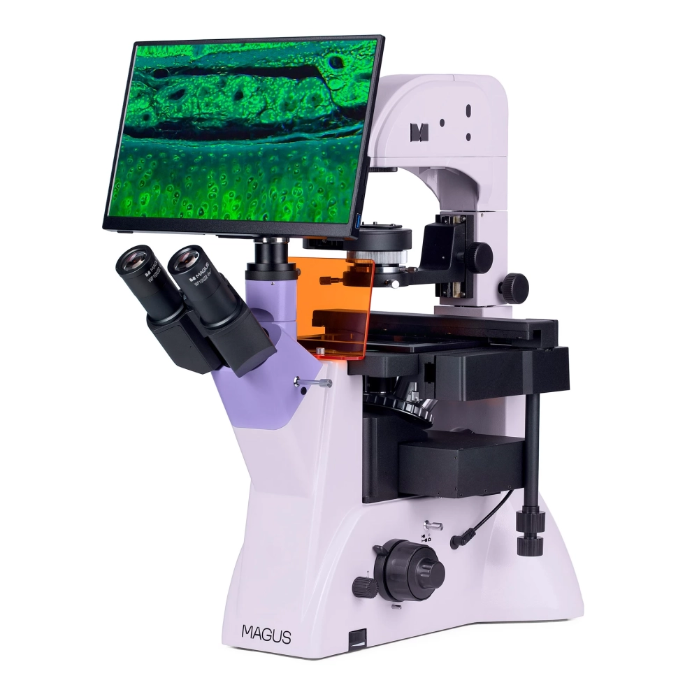
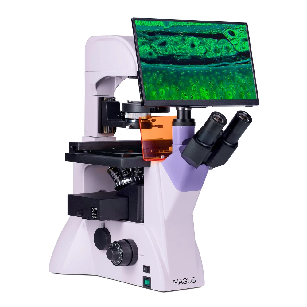
MAGUS Lum VD500L LCD fluorescent inverted digital microscope
Substitute products
Product description
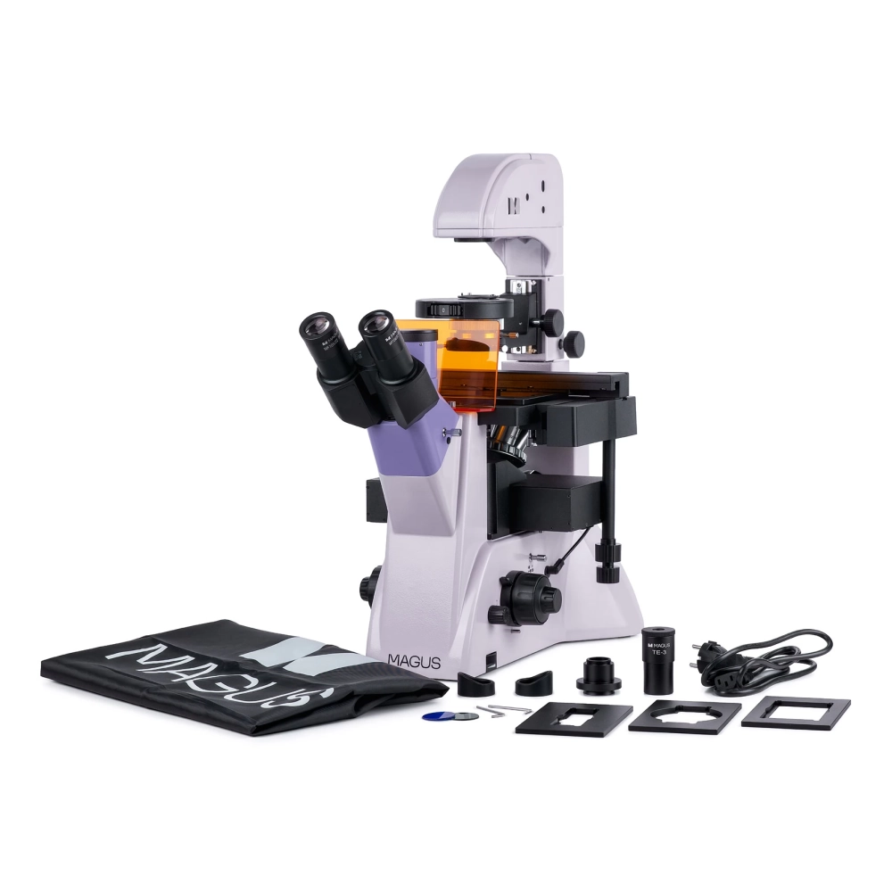
The MAGUS Lum V500L is an inverted microscope for reflected-light fluorescence microscopy and transmitted-light brightfield and phase-contrast microscopy. DAPI, FITC and TRITC fluorochrome filters are used for fluorescence microscopy. The inverted design allows for use in laboratory vessels such as Petri dishes, flasks, etc. observation of existing samples. The height of the laboratory vessels can be 55 mm or 165 mm for tilted stands. The optics are designed for laboratory vessels with a bottom thickness of 1.2 mm. The microscope is great for research and routine laboratory work, as well as for educational purposes.
Digital camera
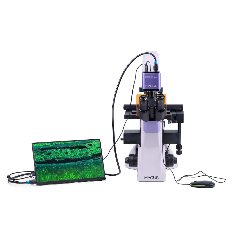
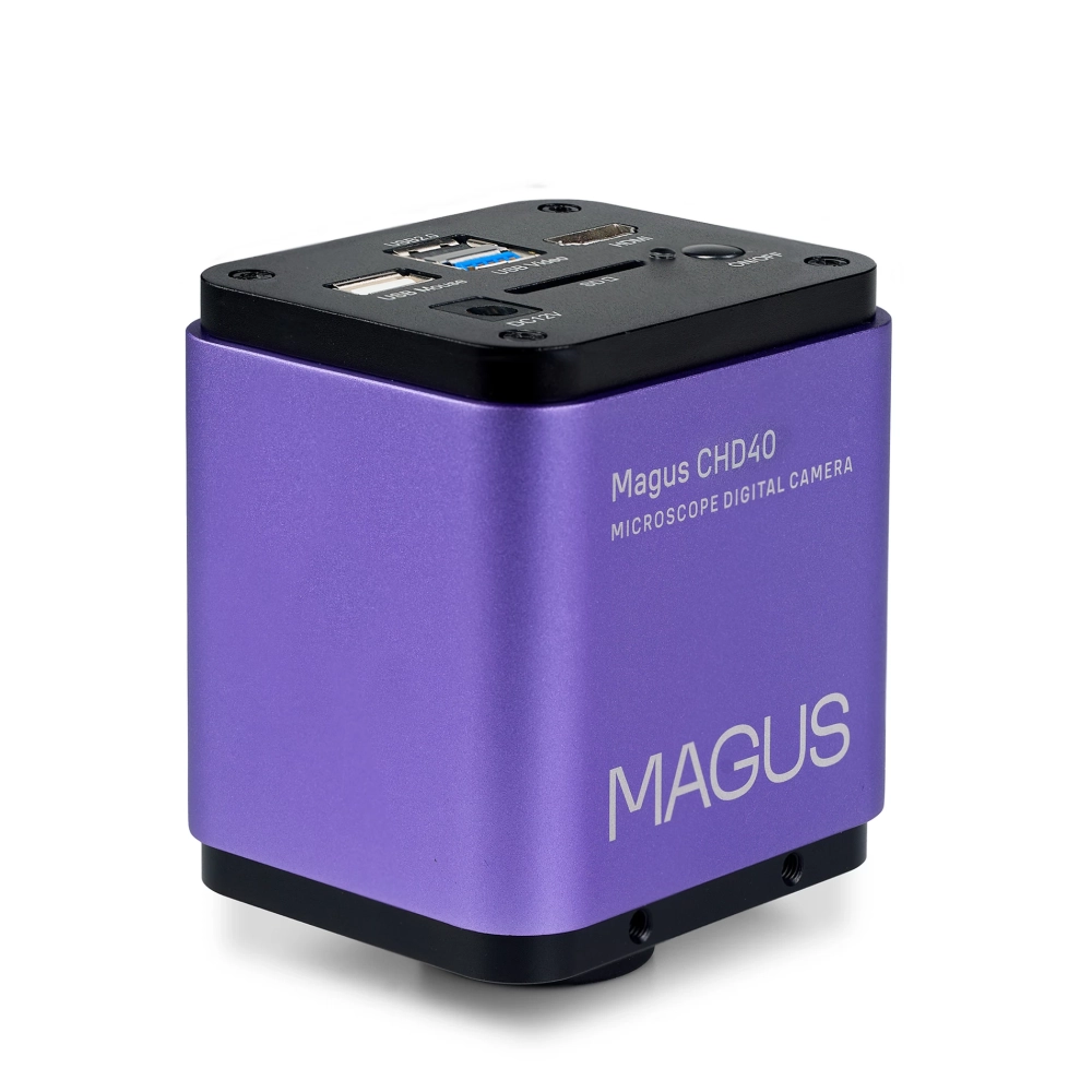
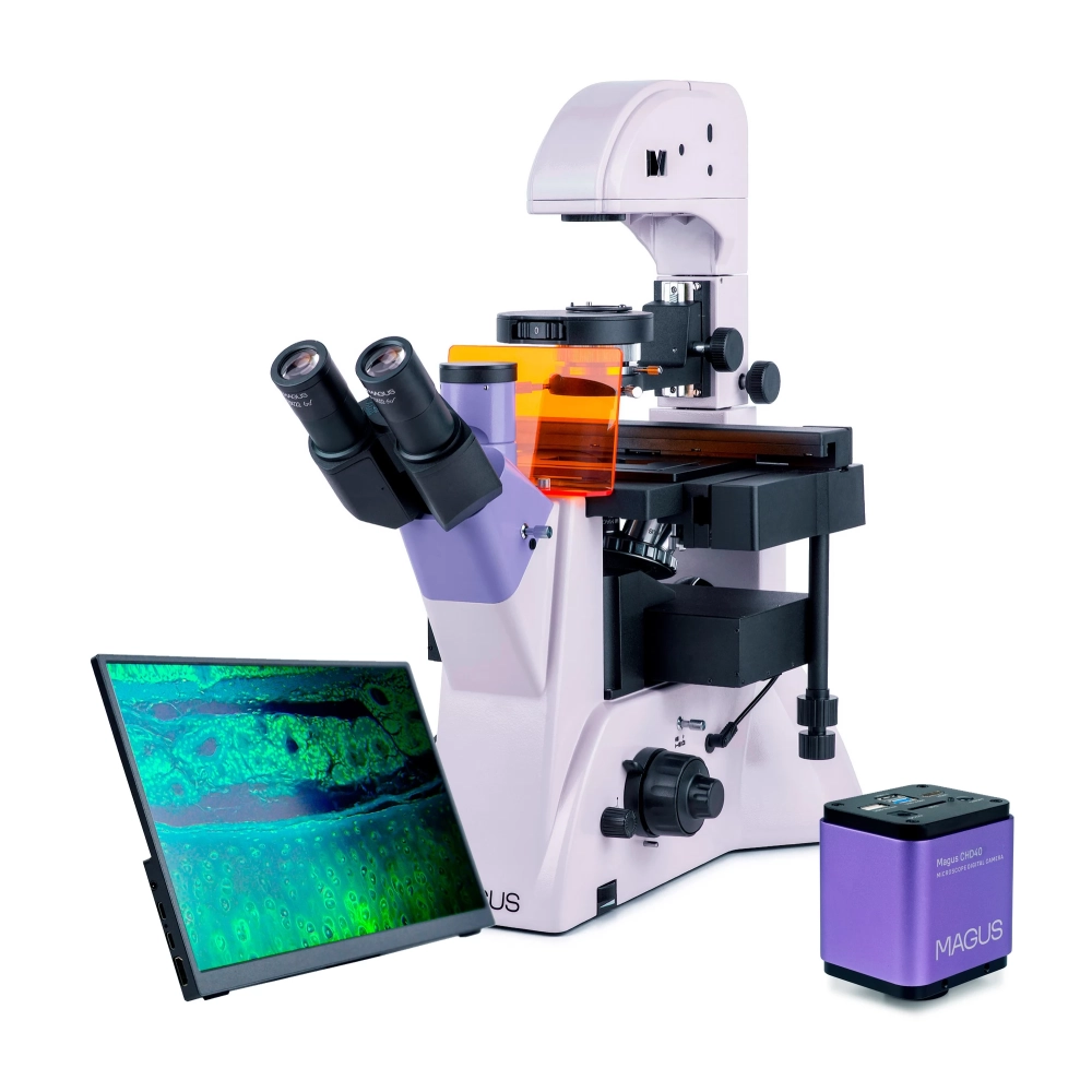
MAGUS CHD40 is a digital HDMI camera with three video interfaces and automatic switching function between 4K and Full HD depending on the monitor resolution.
The camera's 8-megapixel sensor, connected via HDMI or USB 3.0, captures realistic 4K resolution (3840x2160 px) images. When connected via Wi-Fi, Full HD (1920x1080 px) image quality is available. The camera can be connected directly to a TV, monitor or projector with an HDMI interface. In this mode, the camera works independently without a computer. The HDMI connector provides a high and stable transfer rate when transferring from the camera to an external screen. You can connect the camera to a computer via Wi-Fi or USB 3.0. Video is recorded at 30 frames per second (fps).
The camera combines high FPS and high bandwidth HDMI. Thus, the videos have bright colors, there are no freezes or gaps between the frames. At maximum resolution, the image is well detailed, moving objects are visible without error, and the movement of objects is displayed without delay.
Monitor
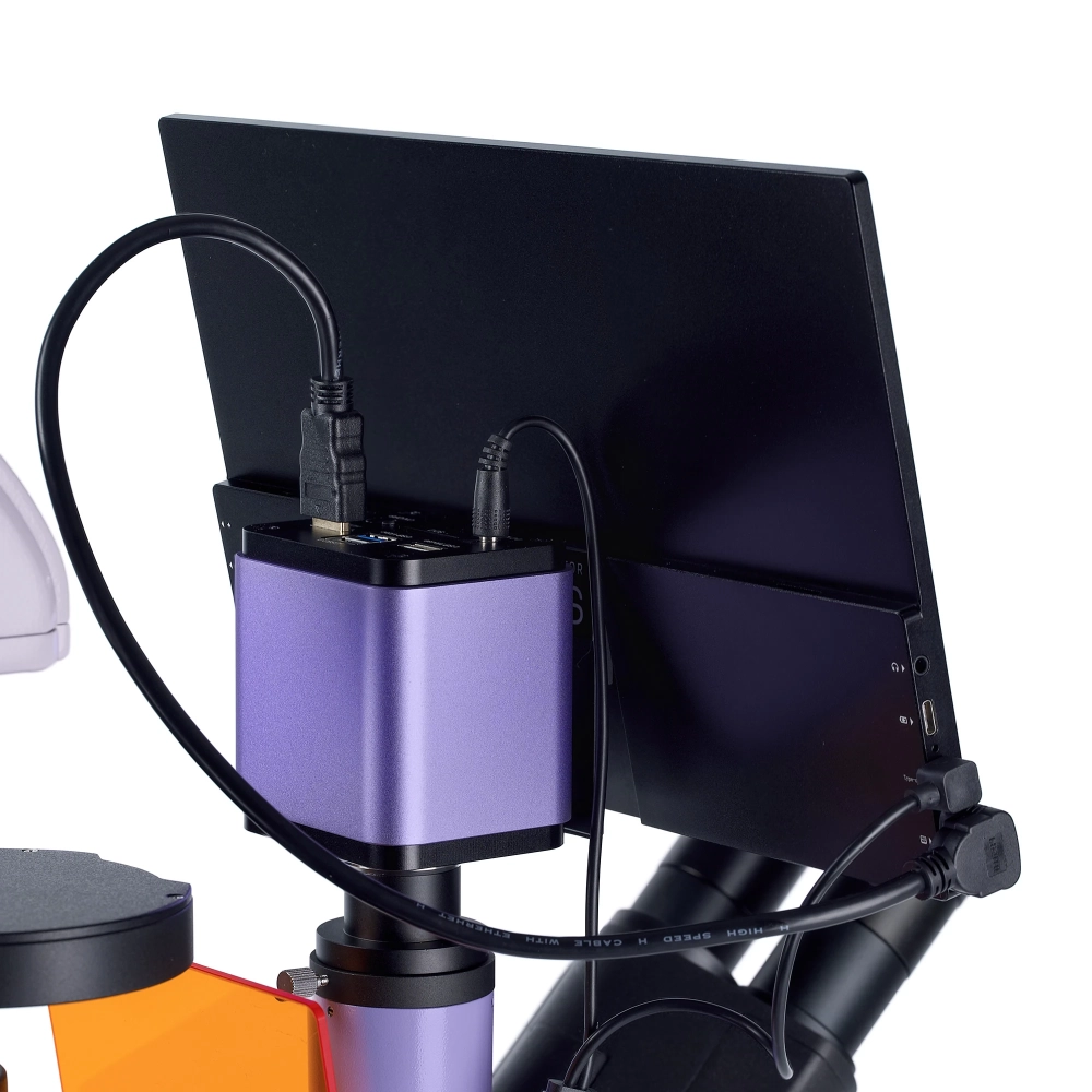
The MAGUS MCD40 monitor is intended for use with the visualization system of the MAGUS microscope. The monitor is connected to a camera mounted on the microscope to display real-time images. MAGUS supports 4K HDMI cameras. The diagonal of the screen is 13.3 inches. The IPS matrix provides bright images with large viewing angles. If you look at the display from an angle, the color reproduction is not distorted. You can place the display on a folding stand, table or shelf, or attach it directly to the camera or microscope stand.
Optical elements
There are two options for mounting a digital camera and monitor: The triple lens tube is used to mount the monitor, and the digital camera can be mounted on the side opening of the frame. There is beam splitting: 100:0/0:100 ratio (on the frame) and 80:20 ratio (on the three-lens head part). Due to the 45° inclination angle of the eyepiece tube, observations can be made in complete comfort. When assembling the microscope, the user can rotate the eyepiece tubes 180°.
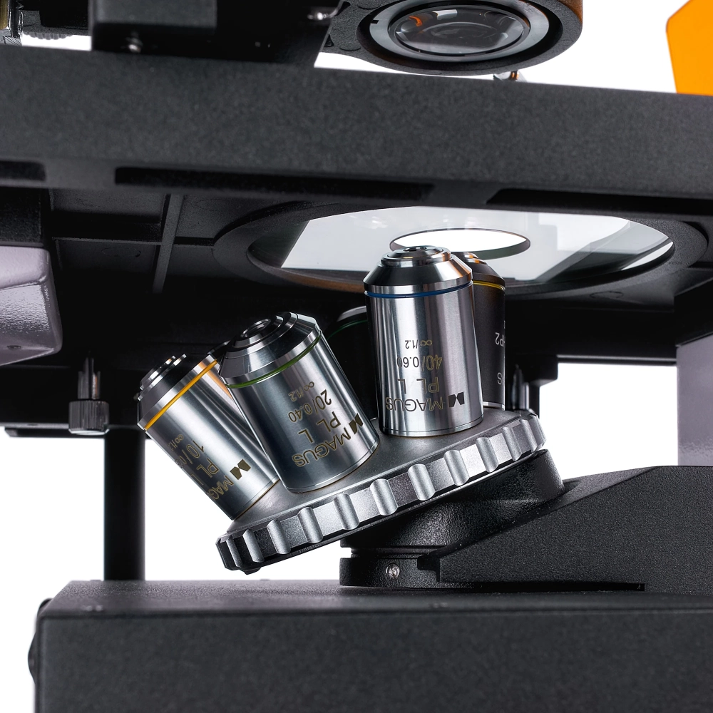
The set includes six planar achromatic objectives: three for brightfield and fluorescence microscopy, and three more for phase contrast microscopy. These are infinity corrected, long working distance lenses. The revolver head has six places to accommodate the objectives. The revolver head is attached to the stand under the specimen table.
Illumination
The fluorescent excitation source is a 5 W LED with a lifetime of 50,000 hours, non-replaceable. There are three excitation filters: DAPI, FITC, and TRITC fluorochrome filters. LED lighting is more energy efficient and easier to maintain than mercury lighting, so it is recommended for use in universities. LED lighting provides uniform colors. The LEDs turn on and off quickly and do not overheat even during long periods of operation.
The source of the transmitted light is also a 5 W LED. Bright enough for bright field and phase contrast microscopy techniques with all objectives.
Both reflected and transmitted illumination can be adjusted according to the Köhler method. This allows the maximum resolution of each objective lens to be achieved and provides a clear, sharp image without artifacts and darkening of the edges.
The microscope has a phase-contrast condenser: Three apertures can be used for 10x, 20x and 40x magnification phase objectives, and the fourth aperture for bright-field microscopy. The phase rings can be centered. You can switch between microscopic techniques by turning the dial.
Stage and focusing mechanism
The fixed stage has a mechanical attachment for moving laboratory vessels and three cup holders of different sizes. The laboratory vessel can be moved in two mutually perpendicular directions in the horizontal plane. The microscope has both a coarse and fine focusing mechanism. The coarse focusing mechanism has a lock button and is adjustable in tension. With the ergonomically designed buttons, the user can work in a comfortable position.
Accessories
The microscope can also be equipped with additional eyepieces, calibration slides or a digital camera from the MAGUS accessory range.
The main characteristics of the microscope
- Reverse construction
NOTEBOOK SEARCH

OPINIONS
0.0
