
Dear Visitor! We are carrying out maintenance this weekend, so our website may be slow or briefly unavailable at times. Thank you for your patience!
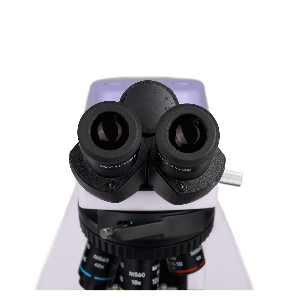
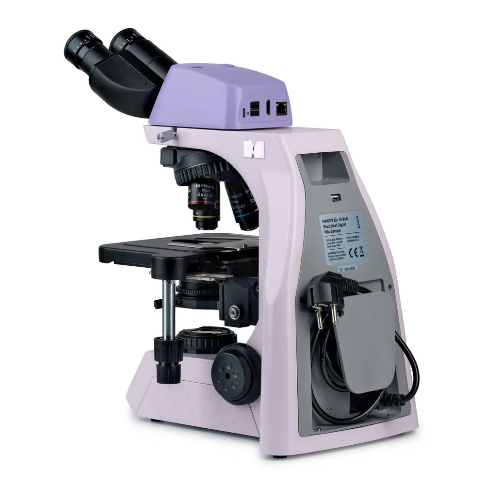
MAGUS Bio DH260 biological digital microscope
1 399 990 Ft
Product description
The microscope is suitable for observing transparent and translucent biological samples, such as smears and cross-sections, using the bright-field microscope technique in transmitted light. By installing additional accessories, the dark field, phase contrast, fluorescent and polarization modes can also be used.
The coded turret maintains comfortable brightness when switching between lenses. The intelligent control of the microscope lighting makes the researcher's daily work more convenient and faster. With the help of "smart" functions, students can find a job in the profession more easily and gain the necessary professional experience.
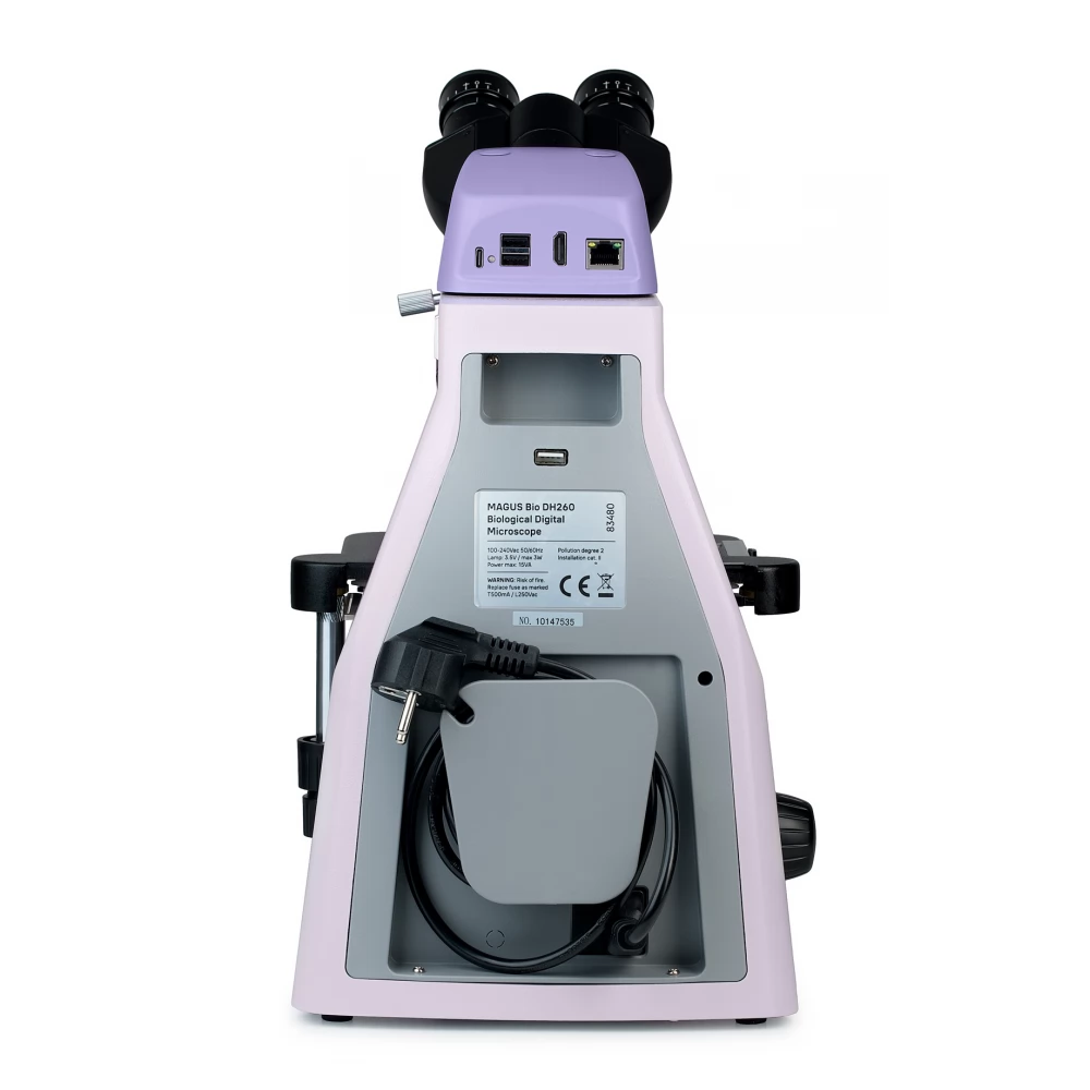
There is a built-in digital camera in the microscope head. The microscope has a minimalist design - no vertical tube or protruding parts.
The microscope is also suitable for laboratory, research and educational work.
Microscope head
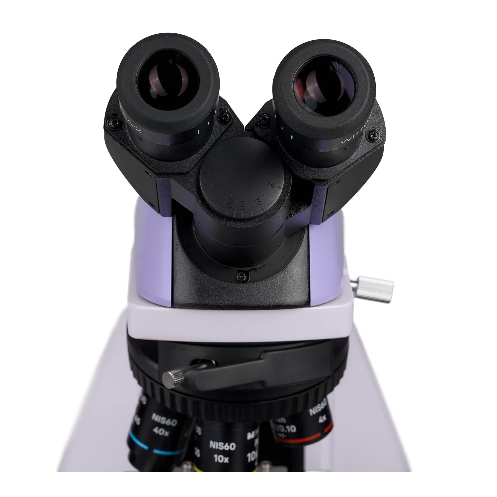
Binocular head part with optical elements corrected to infinity. 360° rotatable eyepiece tube. The user can adjust the eye distance on the eyepiece to match the height.
The basic set includes the 10x/22 mm eyepiece, which can be adjusted to the diopter, and has a large eye distance so that it can be used even with glasses. Flat rubber eyecups protect the optical elements from scratches.
Digital camera
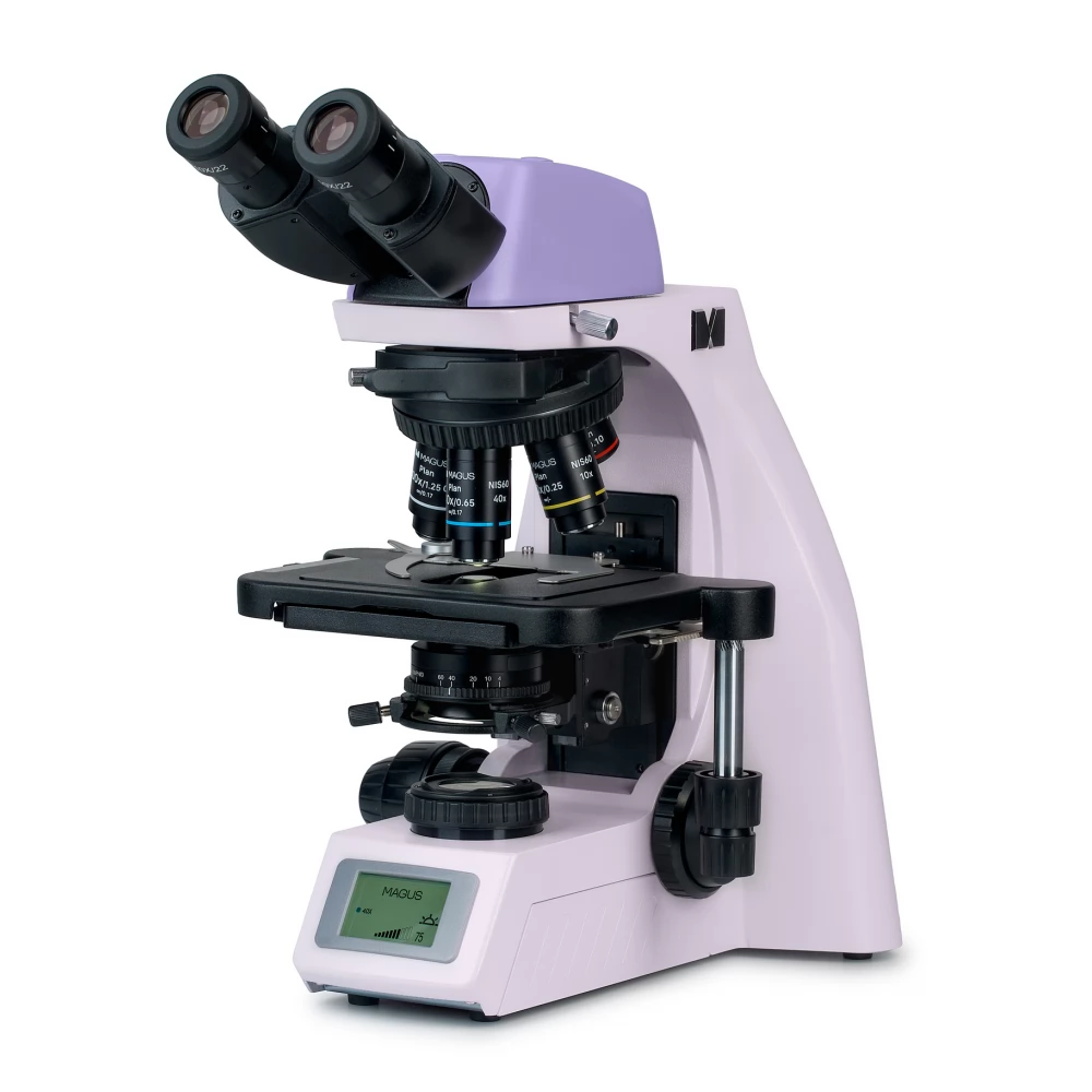
There is a built-in digital camera in the microscope head.
The camera has an 8-megapixel sensor and provides a realistic image with 4K resolution (3840x2160 pixels), which clearly shows the smallest details of the patterns and their structures. The best images can be taken with 4x and 10x lenses.
The video is recorded at maximum resolution at 30 frames per second (fps). The videos are stutter-free, the transition between frames is smooth and smooth. The movement of the sample can be tracked in real time without delay. The camera can be conveniently used for working with moving objects, it is well suited for classroom demonstrations, photographing samples, and fine-tuning the microscope.
With the help of Wi-Fi, you can connect to a computer and transfer data to it.
Turret head and objectives
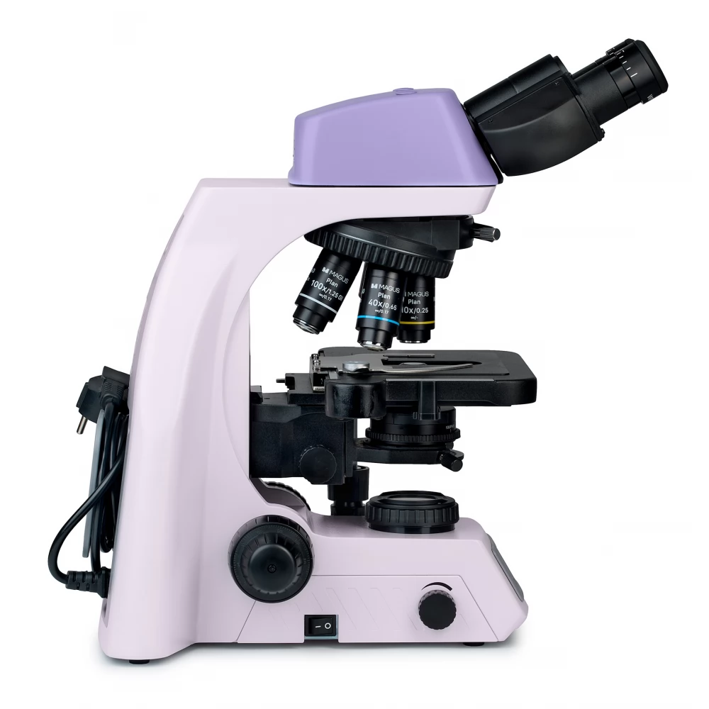
The coded turret holds 5 lenses and faces the inside: the user can see the lens inserted in the optical path, and the area above the stage remains free. An additional lens can be installed in the free aperture to achieve extra magnification. The parfocal distance is 60 mm.
Above the turret head is a hole closed with a plug for installing the analyzer.
Maintaining comfortable brightness when switching between magnifications
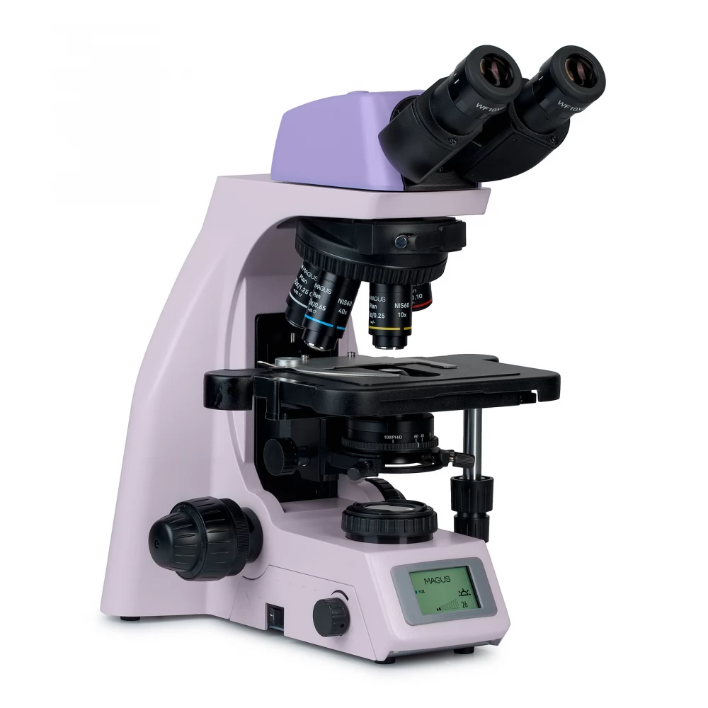
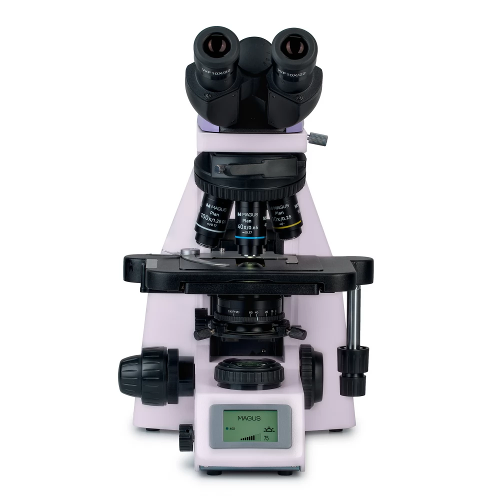
Lenses of different magnification transmit light at different intensities, so when changing lenses, you must always adjust the brightness. Also, the brightness suddenly increases when switching from a higher magnification lens to a lower magnification. The sudden increase in brightness causes eye fatigue. MAGUS Bio DH260 has intelligent brightness control. The microscope remembers the brightness for each objective selected by the user and automatically adjusts the brightness as the turret is turned. Intelligent control reduces the time required to adjust the brightness. The MAGUS Bio DH260 makes work more convenient and saves time, even if you often have to change the magnification during work.
Focusing mechanism
Coaxial coarse and fine focus adjustment buttons.
The coarse focus adjustment knob is on the left side. The fine focus adjustment buttons are located on both sides; the right button has recesses for the fingers.
The coarse focus lock button allows quick adjustment of the microscope after changing the test object. The knob is located on the left side of the microscope, on the same axis as the focusing mechanism.
The ring on the right adjusts the tightness of the coarse focus range of motion. The user can set a voltage that is comfortable for work.
Object table
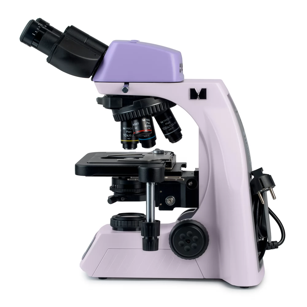
In order to ensure the ergonomic operation of the specimen table, we did not place an X-axis adjustment stand on it. The belt-driven structure enables smooth movement of the pattern. The sample holder is fixed by two screws, which can be easily removed during manual scanning.
The long stage control handle ensures the user's comfort while working: the head of the hand can rest on the table without tiring.
Condenser
The condenser is adjustable in height and can be centered. Fastening type: dovetail.
The condenser ring adjusts the iris aperture. On the body of the condenser there is a marking for the magnification of the objectives, and on the ring there is an indicator marking. To achieve contrast on individual lenses, it is recommended to turn the ring so that the reference mark corresponds to the magnification mark of the lens being used.
The condenser has a slot with a plug for the dark field or phase contrast slider. The slider saves time when changing the monitoring method.
Illumination
The transmitted light is provided by a 3 W LED. The color temperature does not change
PRICE NOTIFICATION
NOTEBOOK SEARCH

OPINIONS
0.0
