
Dear Visitor! We are carrying out maintenance this weekend, so our website may be slow or briefly unavailable at times. Thank you for your patience!
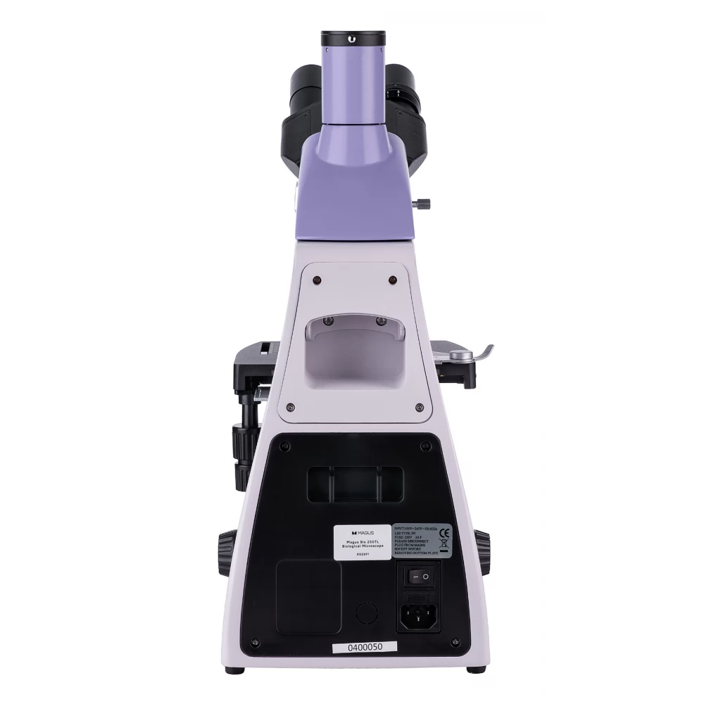
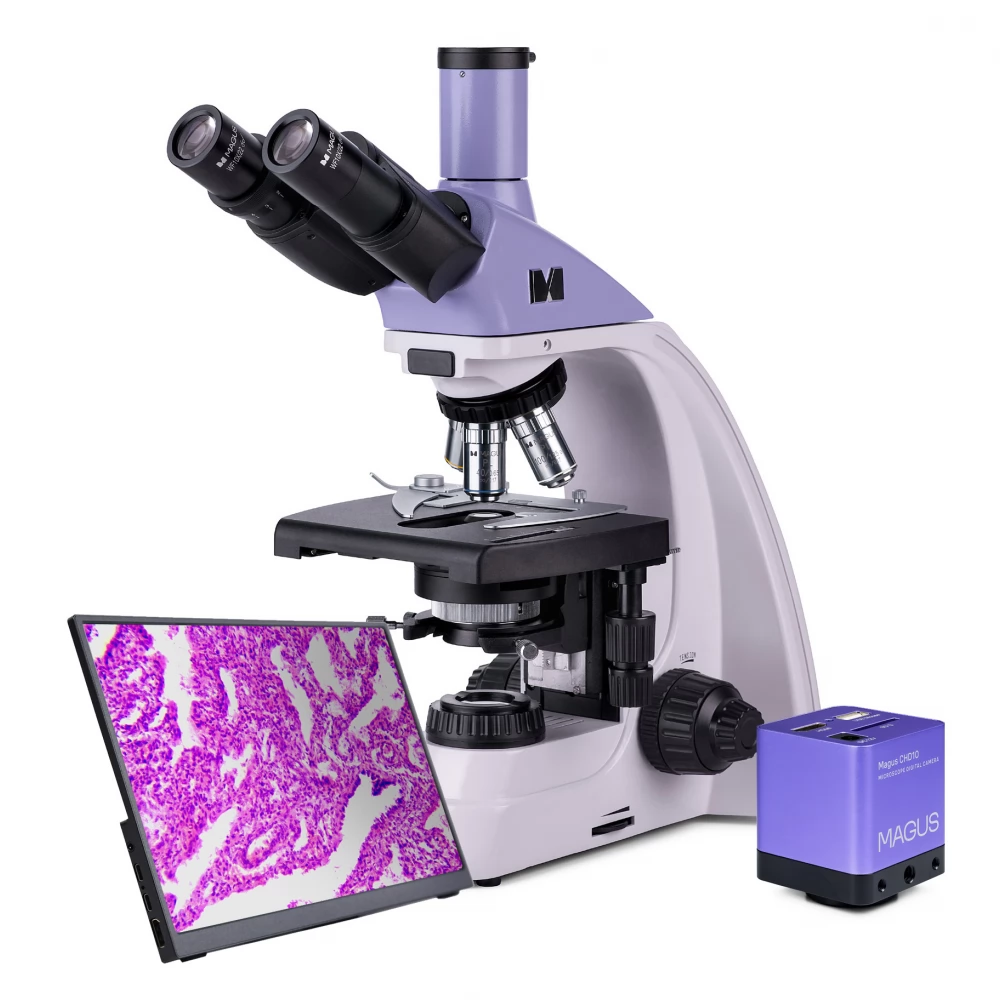
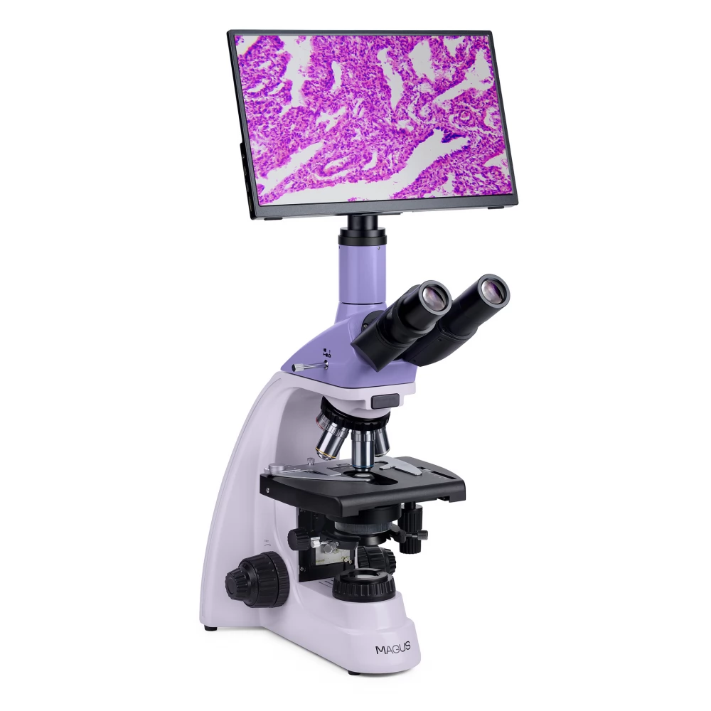
MAGUS Bio D250TL LCD biological digital microscope
889 989 Ft

Product description
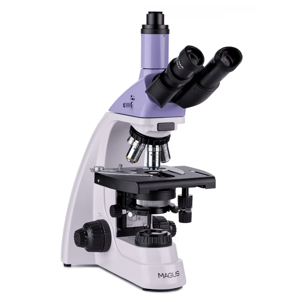
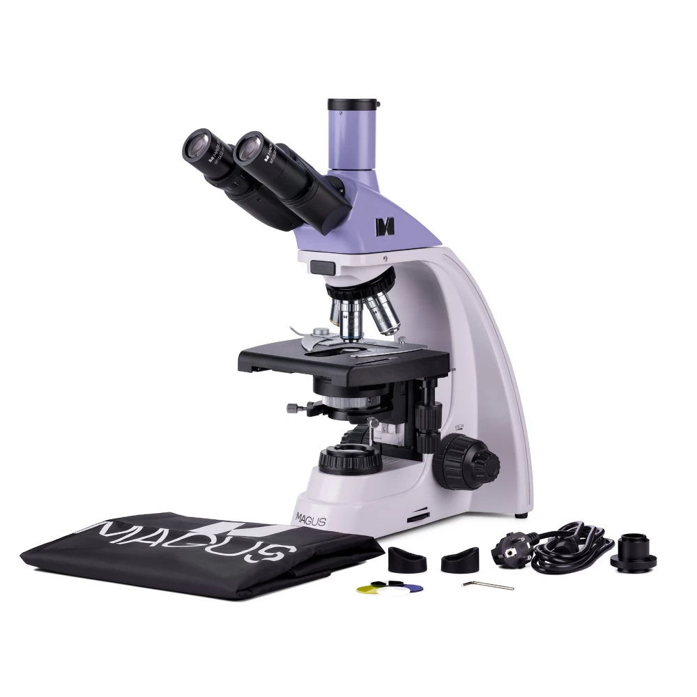
With the MAGUS Bio D250TL LCD microscope, thin sections and smears of biological samples can be studied. The observation is carried out using a bright-field microscope technique with transmitted light. The light source is a 3-watt LED, and the optics consist of infinite plane achromatic lenses. The microscope supports additional accessories, including darkfield, polarization and phase contrast instruments. The microscope is an ideal choice for laboratory analysis, research and teaching.
Digital camera
The MAGUS CHD10 camera is equipped with a 2 MP sensor, so it captures realistic Full HD images with a resolution of 1920x1080 pixels.
It is an autonomous camera that does not need to be connected to a computer and does not require software installation. The camera can be connected directly to a TV, monitor or projector to display the image. The HDMI connector provides a high and stable transfer rate when transferring from the camera to an external screen. It records video at 60 fps.
The camera combines high FPS and high bandwidth HDMI. Thus, the videos have bright colors, there are no freezes or gaps between the frames. At maximum resolution, the image is well detailed, moving objects are visible without error, and the movement of objects is displayed without delay.
Monitor
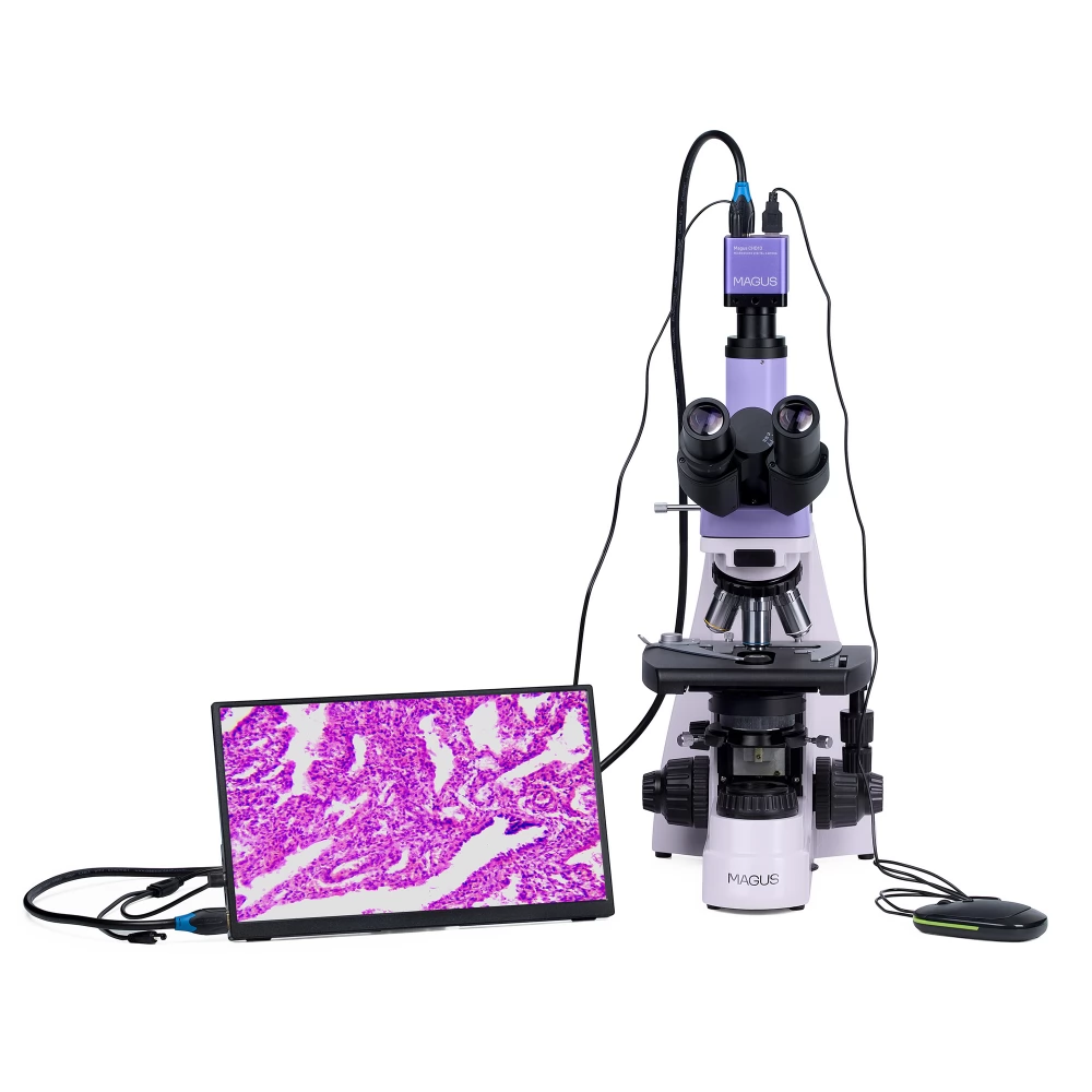
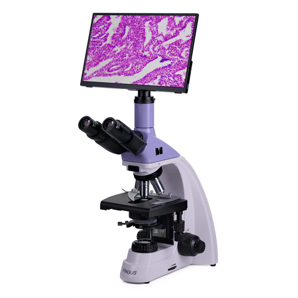
MAGUS MCD monitors combined with MAGUS CHD cameras provide a complete imaging system for MAGUS microscopes.
The camera is connected via the HDMI interface. The monitor displays a real-time image. Its IPS LCD display provides a wide viewing angle, high brightness and good contrast. The image is not distorted, even if we look at the screen from a wide angle. The screen size is 13.3''.
The monitor can be installed on a horizontal surface using a foldable stand, or can be attached directly to the camera or tripod.
Optical elements
The three-eyepiece head part includes two eyepiece tubes for observations and a vertical tube for attaching the digital camera (the camera is not included). The tubes have an angle of inclination of 30°, so that they can be used comfortably even when working for long periods of time, and they can be rotated 360° so that they can be adjusted to the height of the observer. There is a diopter adjustment ring on the left tube.
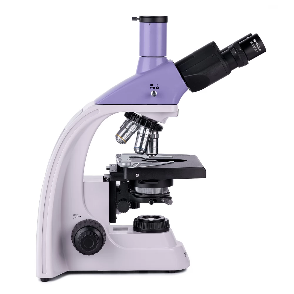
The lenses are mounted in a five-slot turret – four lenses are attached, while one slot is left free for an additional lens. Any optional objective lens can be mounted in the open aperture, allowing additional magnification within the zoom range. The user can see the lenses inserted into the optical path as the turret faces the inner part. This leaves the area above the specimen table free.
The magnification range can be set between 40x and 1000x (only valid for the basic configuration). By adding additional lenses, the upper magnification limit can be increased.
Illumination
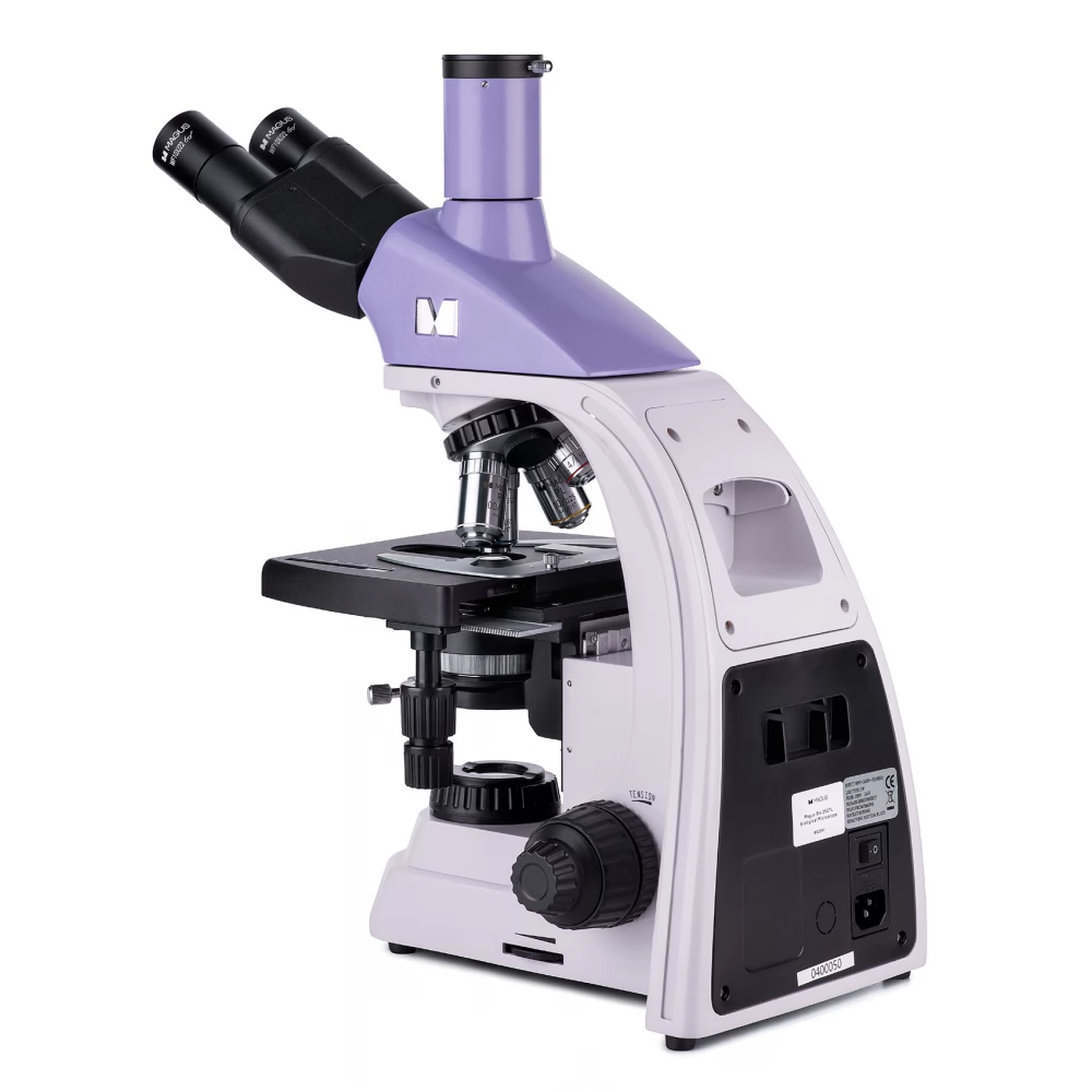
LEDs mounted on the bottom of the microscope ensure bright and even illumination of the object. The color temperature is not changed by changing the brightness. The power of the light fixture is 3 W. Its lifespan is 50,000 hours. The lighting system is powered by an AC power supply.
By adjusting the position of the Abbe condenser and by opening/closing the iris and field diaphragms, it is possible to adjust the Köhler illumination. For dark field and phase contrast techniques, you can use sliders (they have a special opening in the condenser). Switching between research techniques is extremely fast with the slider.
Stage and focusing structure
The specimen table does not have a positioning stand. Thus, it does not interfere during observations, and the microscope itself takes up less space on the table. The mechanical mount that moves the sample can be removed (which is useful for manual scanning).
The focus can be adjusted using coarse and fine focusing. The buttons are located on both sides of the microscope. Since they are located closer to the sole, you don't have to keep your hands in the air while working. You can also calm them down on the table. The coarse focus has a lock button and is adjustable in tension. Focus can be adjusted smoothly and effortlessly.
The main characteristics of the microscope
- Three-eye lens head, turret head for 5 lenses, infinite plane achromatic optics
- The eyepiece tubes can be rotated 360° for easy adjustment to the height of the observer
- There is a diopter adjustment ring on the left tube, coarse and fine focusing
- The coarse focus has a locking button and its tension can be adjusted
- The Abbe condenser is centralized and adjustable in height, and the field and iris diaphragms allow Köhler illumination to be adjusted
- Transmitted light source 3 W LED with adjustable brightness, operated by a 220 V power supply
- The possibility of installing additional accessories
Main features of the camera
- The camera works independently, without connecting to a PC or installing any software
- HDMI interface for high and stable data transmission speed
- The camera
PRICE NOTIFICATION
NOTEBOOK SEARCH

OPINIONS
0.0
