
Dear Visitor! We are carrying out maintenance this weekend, so our website may be slow or briefly unavailable at times. Thank you for your patience!
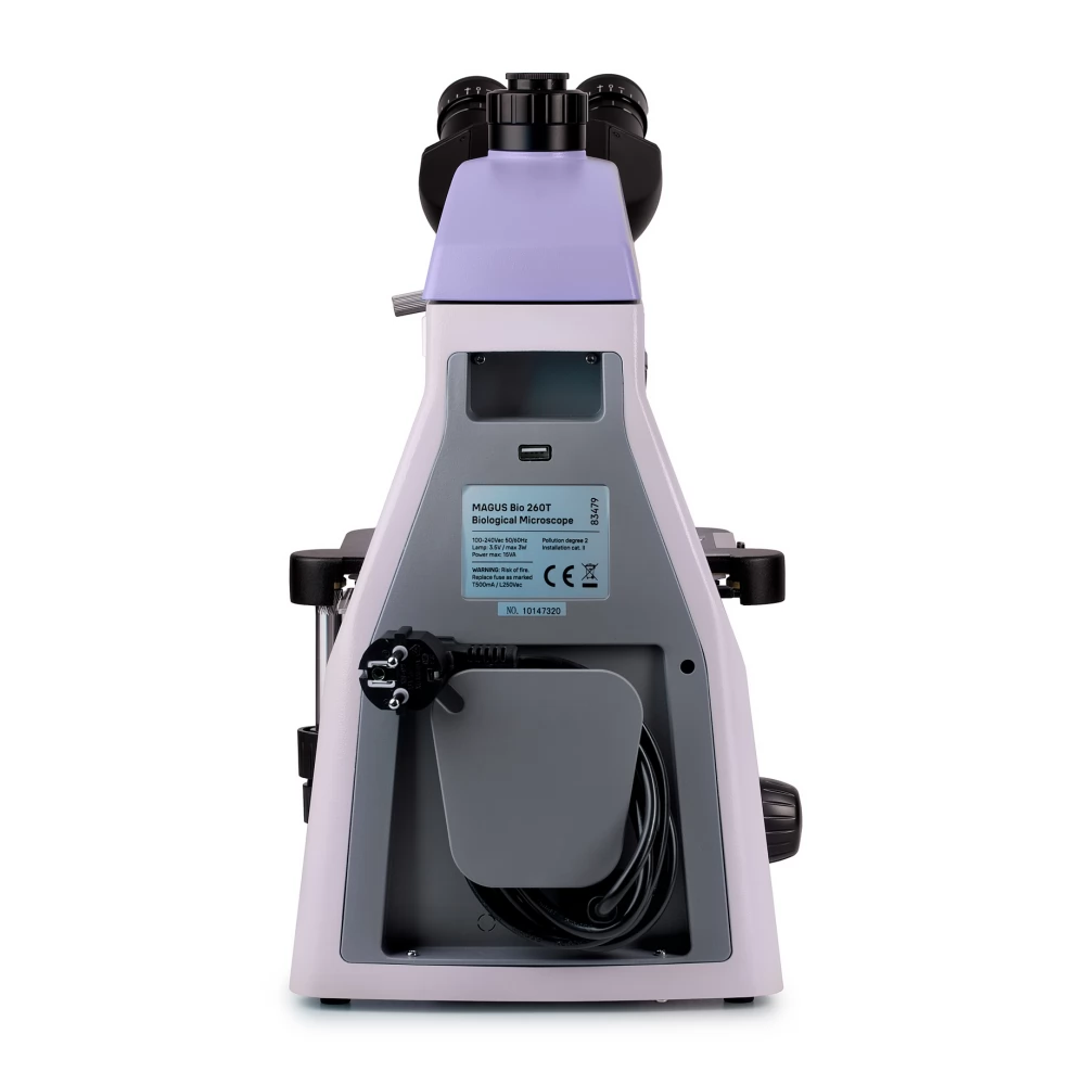
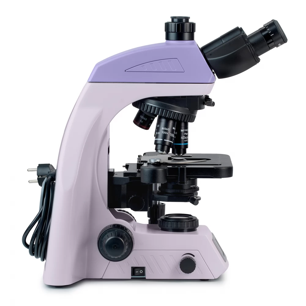
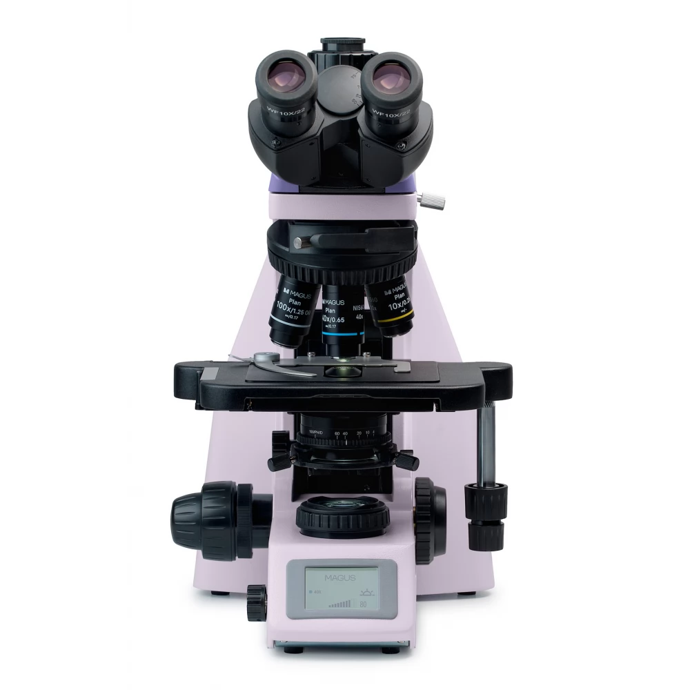
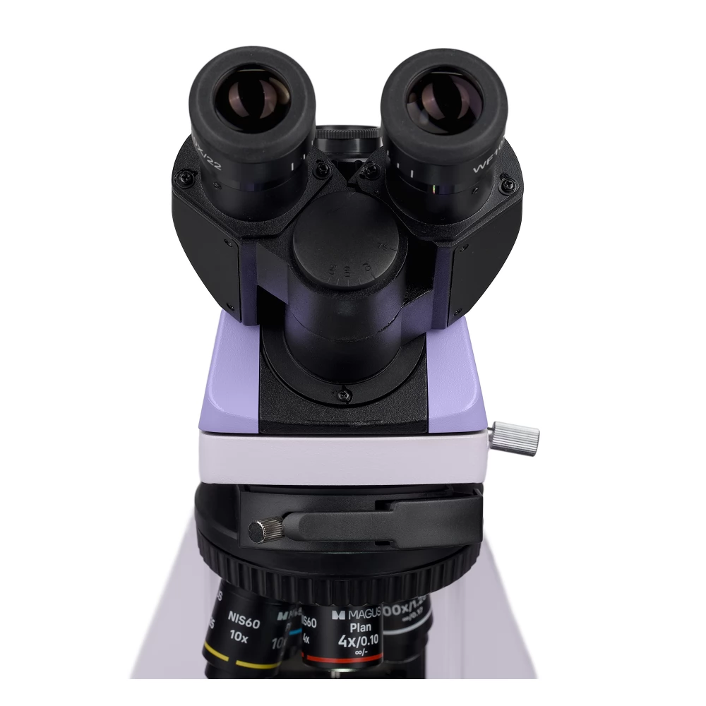
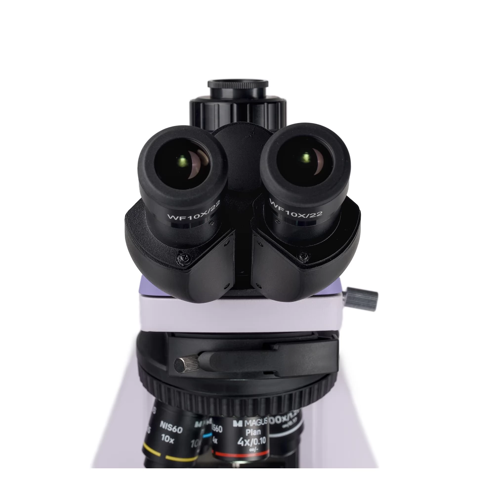
MAGUS Bio 260T biological microscope
999 989 Ft

Product description
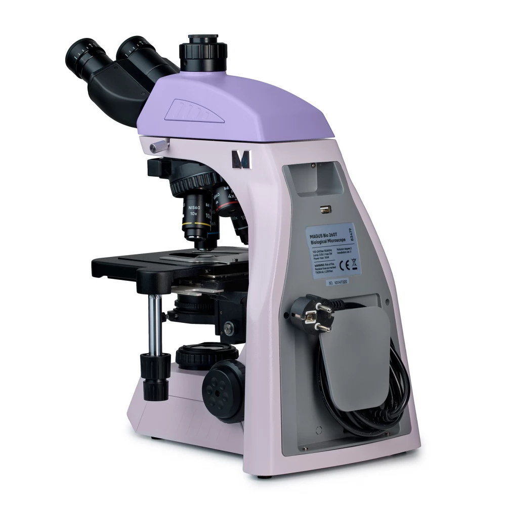
The MAGUS Bio 260T model is a microscope that can be used for laboratory and scientific research or for educational purposes in everyday use. With the microscope, thin, transparent and translucent samples can be studied using the bright field method. It can also be equipped with additional components, so the possibilities of the equipment can be extended to dark field, phase contrast, polarized and fluorescent lighting methods.
The model has a coded turret that can accommodate up to five lenses. By rotating the turret, the "intelligent" lighting switches automatically and adjusts to the parameters of each object. Thanks to this function, you don't have to waste time adjusting the light. This can be particularly important in the teaching process or if the magnification value needs to be changed frequently.
The three-eye lens head of the microscope works with an optical system corrected to infinity. The pupil height can be adjusted. Its position can be adjusted by rotating the tubes 360°. A digital camera can be placed in the triple-lens tube. Light enters the camera and the eyepieces in a 50/50 ratio. The microscope set includes 10x/22 mm basic eyepieces with a diopter adjustment mechanism. The tubes are not adjusted. The rubber eyecups included in the set are beneficial for users who wear glasses, because they protect the glasses from wear and scratches.
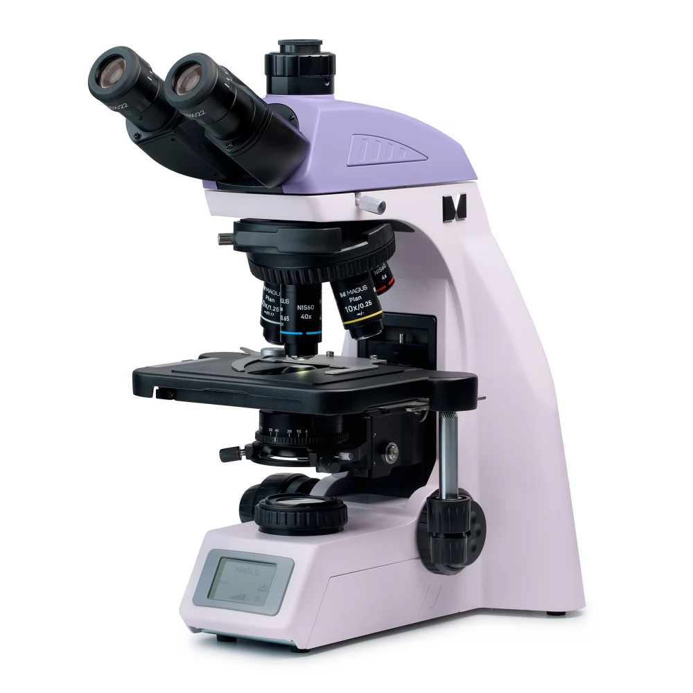
The rotating turret has a 5-lens automatic lighting control system. When changing the magnification, the user notices a strong fluctuation in the level of brightness in the eyepieces: lenses with different magnification values allow light to pass through in different ways. Intelligent lighting allows you to set the light level for a particular lens once, then turn the turret, and the equipment will automatically adjust the light. There are 4 objectives in the microscope set. The user can even place a lens in the fifth aperture if desired. The parfocal distance is 60 mm. The opening above the rotating turret head is used to fix the analyzer. If the analyzer is not installed, the opening must be closed with a plug.
The specimen table was designed with a belt-driven mechanism: so the sample moves smoothly and evenly. Working with the microscope is more ergonomic and comfortable because there is no X-axis positioning stand. The sample holder is fixed by two screws, which can be easily removed if necessary. Another convenience feature is the long stage control arm. The user can use the microscope with his hands resting on the table without straining his arms or body.
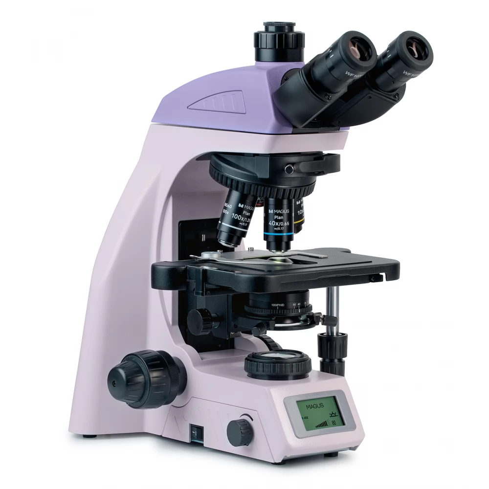
The condenser can be adjusted to the desired position both vertically and relative to the optical axis (fixed on a curved support). The iris diaphragm can be adjusted with a ring: there is a mark on it, and the magnification marks of the objective can be seen on the condenser frame. In the case of a contrasted image, the indication must be set according to the installed work objective. The condenser can also be supplemented with dark-field or phase-contrast slides: for this, the equipment is equipped with a special opening.
The microscope can be focused with the coarse and fine adjustment buttons located coaxially on it. Coarse focusing can be done with the left button. The fine focusing buttons are located on both sides of the tripod. The one on the right is specially grooved for the comfort of the fingers. The coarse focus adjustment clamp and the range of motion adjustment ring provide additional convenience: after changing the lens, you can use the button to adjust the focus, and the ring allows you to adjust the tightness of the coarse focus range of motion to your liking. Both the buttons and the ring are located at the bottom of the stand: the user can control them while resting his hand comfortably on the table.
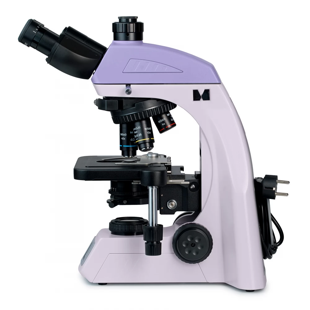
The microscope is illuminated by a 3 W LED installed in a transmitted light fixture. Even when the brightness changes, the lighting temperature remains stable. The microscope's optional research methods include Köhler illumination. As a result, the entire sample field is uniformly illuminated, and no dust or light source image will be visible through the eyepiece or digital camera.
The selected operating parameters can be viewed and set on the built-in display. It is located at the bottom of the microscope. The display shows the magnification value of the working lens, the lighting parameters, the operating mode and the automatic switch-off time.
The functionality of the device can be expanded with additional elements. The magnification spectrum can be increased with a set of eyepieces. For additional methods of working with samples that cannot be seen by the bright field method, the microscope must be supplemented with a phase contrast device, a dark field slide or a polarizing device. With the help of the digital camera, the results of the research can be recorded on a photo or video, or the image can also be displayed on the screen. The objects
PRICE NOTIFICATION
NOTEBOOK SEARCH

OPINIONS
0.0
