
Dear Visitor! We are carrying out maintenance this weekend, so our website may be slow or briefly unavailable at times. Thank you for your patience!
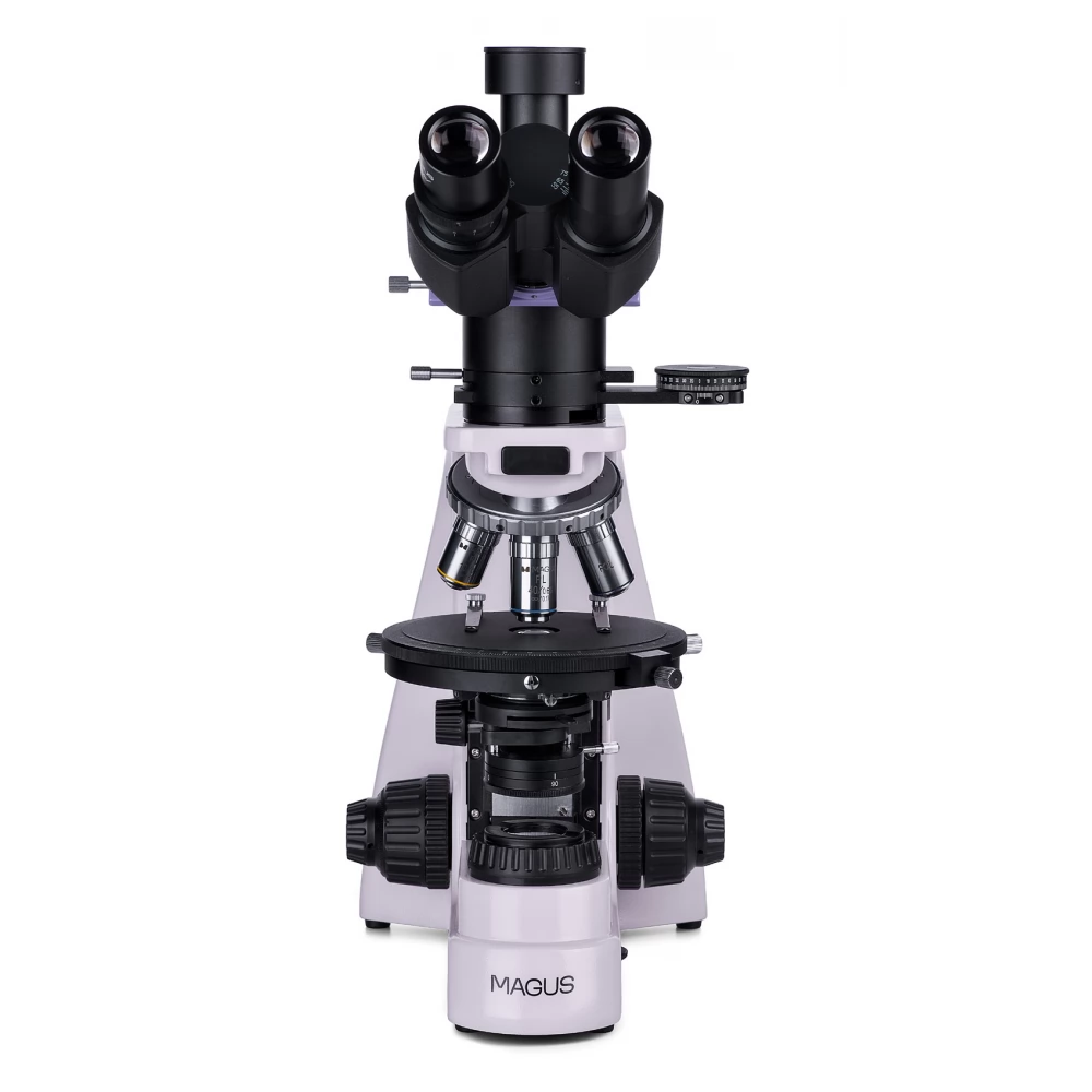
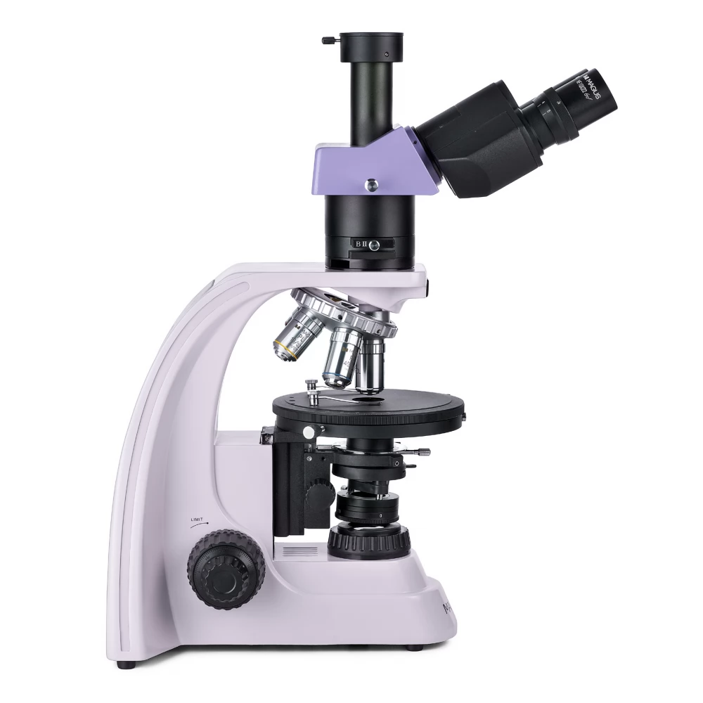
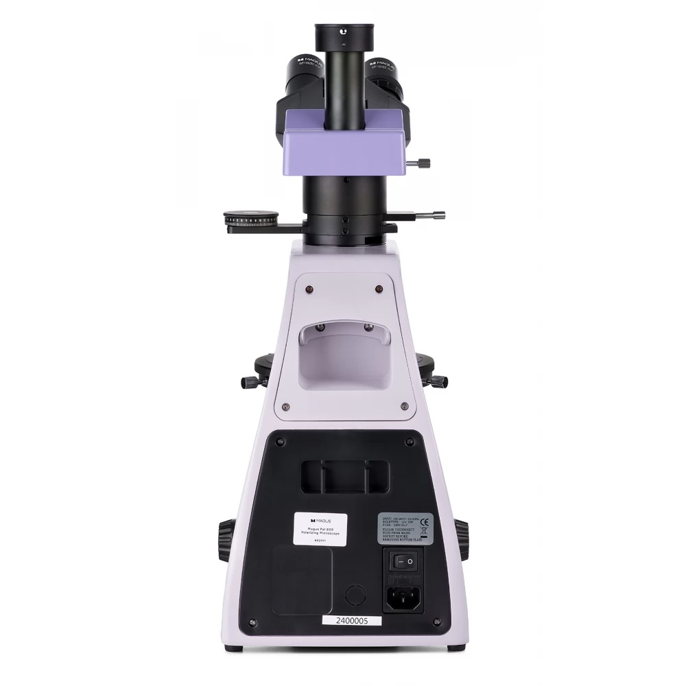
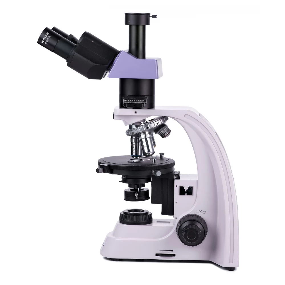
MAGUS Pol D800 LCD polarizáló digital microscope
1 899 990 Ft
Product description
The MAGUS Pol 800 polarizing microscope is used to study anisotropic objects in polarized and ordinary transmitted light. The objects to be studied can be of biological and geological origin, or polymers. The optical elements of the microscope are voltage-free, i.e. they do not create disturbing light refractions. They create a clear, contrasting, authentic image. The functions of the microscope can be used in many fields of science: medicine, forensics, mineralogy, rock and crystal science, etc.
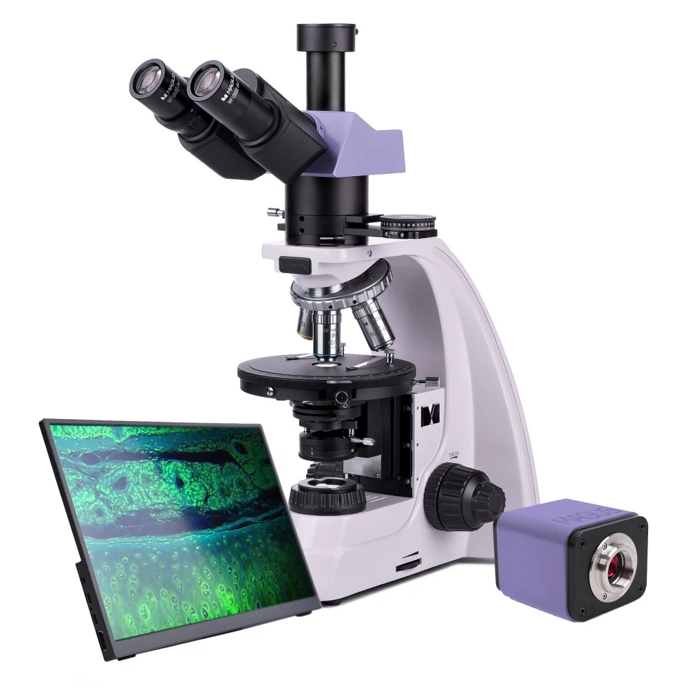
Digital camera
MAGUS CHD40 is a digital HDMI camera with three video interfaces and automatic switching function between 4K and Full HD depending on the monitor resolution.
The camera's 8-megapixel sensor, connected via HDMI or USB 3.0, captures realistic 4K resolution (3840x2160 px) images. When connected via Wi-Fi, Full HD (1920x1080 px) image quality is available. The camera can be connected directly to a TV, monitor or projector with an HDMI interface. In this mode, the camera works independently without a computer. The HDMI connector provides a high and stable transfer rate when transferring from the camera to an external screen. You can connect the camera to a computer via Wi-Fi or USB 3.0. Video is recorded at 30 frames per second (fps).
The camera combines high FPS and high bandwidth HDMI. Thus, the videos have bright colors, there are no freezes or gaps between the frames. At maximum resolution, the image is well detailed, moving objects are visible without error, and the movement of objects is displayed without delay.
Monitor
The MAGUS MCD40 monitor is intended for use with the visualization system of the MAGUS microscope. The monitor is connected to a camera mounted on the microscope to display real-time images. MAGUS supports 4K HDMI cameras. The diagonal of the screen is 13.3 inches. The IPS matrix provides bright images with large viewing angles. If you look at the display from an angle, the color reproduction is not distorted. You can place the display on a folding stand, table or shelf, or attach it directly to the camera or microscope stand.
Optical elements
There is a three-eye tube on the three-eye lens head for mounting a digital camera. Using the switch on the frame of the device, the light can be directed entirely to the eyepiece tube or the digital camera. Due to the 30° inclination angle of the eyepiece tubes, long-term observation is also more comfortable, the neck muscles do not get tired. On the microscope, the optics of the diopter adjustment ring of the left tube (on which the tube rotates) can be adjusted to the unique vision of the user.
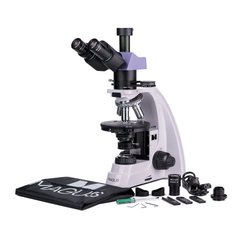
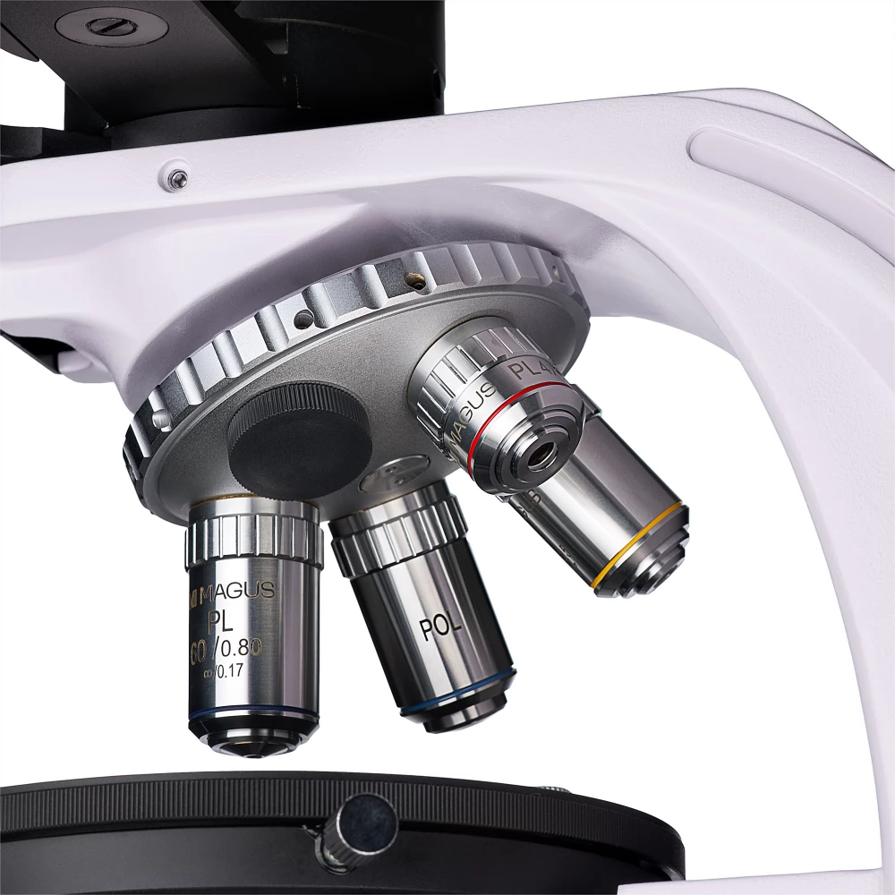
The turret head was designed in such a way that five different lenses can be inserted into it at the same time. There are four of them in the package. Additional lenses can be purchased separately from the MAGUS accessory selection. The lenses can be used with a cover plate with a thickness of 0.17 mm. Thanks to the "away from the observer" turret, the lens inserted in the optical path is clearly visible during work, so the researcher can work more comfortably. The grooves of the revolver head are located in the middle.
Illumination
The light source is located under the object table, which means that the observation can be carried out in transmitted light. The 30 W halogen bulb provides strong, but eye-friendly illumination that can be used with all lenses. The illumination adjusted using the Köhler method results in stronger contrast and better image quality.
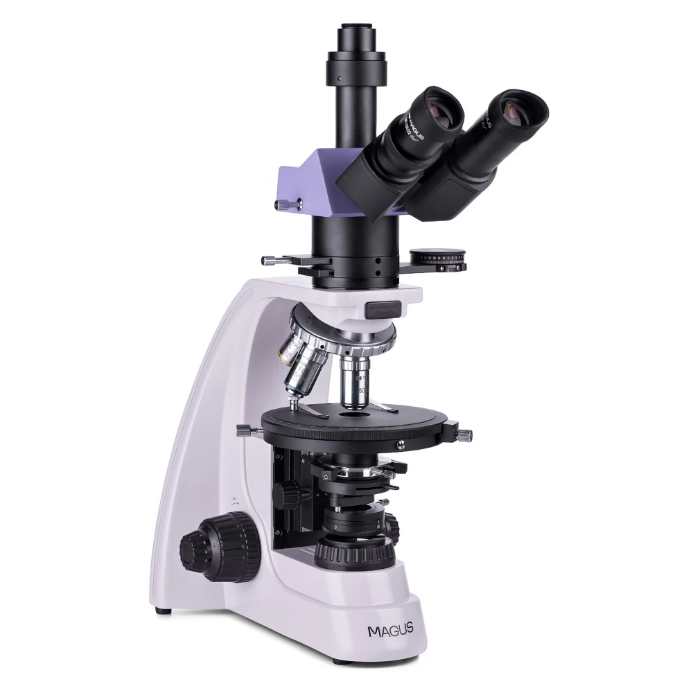
The microscope is equipped with a polarizer and the analyzer. To work in polarized light, the analyzer is located in the optical path. The polarization angle can be changed by rotating the polarizer and the analyzer relative to each other. On the microscope, the Bertrand lenses are held by a central assembly, and this assembly also has the grooves for the compensators. Compensators improve the contrast of low birefringence samples. Bertrand lenses can be used for conoscopic examinations.
Stage and focusing mechanism
The stage of the microscope rotates, and this allows rapid changes in the refraction of the specimen when working in polarized light. The stage is located in the middle of the optical axis of the microscope, the rotation angle can be adjusted gradually, and the tilt angle can be determined with an accuracy of 0.1° using the scale.
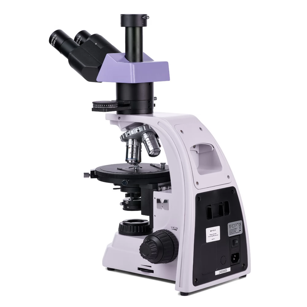
There is a coarse and a fine focus adjustment element on the focusing mechanism. The coarse focus adjustment latch can be used to adjust the coarse focus, and the coarse tension can also be adjusted for comfortable observation.
Accessories
Additional eyepieces, objectives, digital cameras and calibration slides can be purchased for the microscope. With these accessories, the functions of the microscope applicable in research areas can be expanded.
- Observations in transmitted light, in a clear field of view, and with the polarization method
- The lighting
PRICE NOTIFICATION
NOTEBOOK SEARCH

OPINIONS
0.0
